
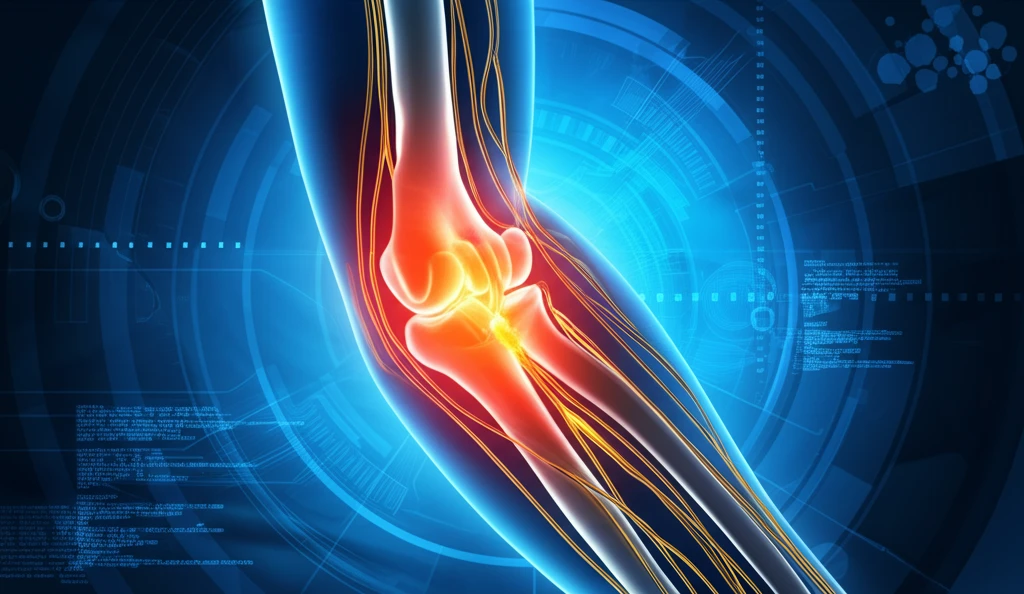
Unlocking Elbow Pain Relief: Surgical Insights into Nerve Compression

"Exploring innovative surgical techniques to alleviate median nerve compression at the elbow and restore functionality."
Elbow pain, often dismissed as a minor inconvenience, can significantly impact daily life, especially when caused by median nerve compression. This condition, characterized by pressure on the median nerve as it passes through the elbow, can lead to pain, numbness, tingling, and weakness in the forearm and hand. Understanding the causes, diagnosis, and treatment options for median nerve compression is crucial for those seeking lasting relief.
While conservative treatments like physical therapy and medication can provide temporary relief, surgical intervention may be necessary in more severe cases. Recent advancements in surgical techniques offer promising solutions for decompressing the median nerve and restoring functionality. One such technique involves the isolated section of the lacertus fibrosus, a fibrous band that can contribute to nerve compression.
This article delves into the surgical approach for treating median nerve compression at the elbow, focusing on the innovative technique of sectioning the lacertus fibrosus and highlighting the importance of accurate diagnosis and comprehensive care for individuals suffering from this debilitating condition.
The Median Nerve and Clinical Burden of Compression
The median nerve is one of the major peripheral nerves of the upper limb, originating from the brachial plexus and serving both motor and sensory functions throughout the arm. The median nerve and its branches are susceptible to compression at the elbow and forearm, producing characteristic patterns of motor and sensory deficits. Clinical presentations of these entrapment syndromes frequently overlap, and electrodiagnostic tests are often inconclusive, complicating accurate diagnosis.
Diagnostic Testing and Its Accuracy Gaps
Hand nerve tests assess the median, ulnar, and radial nerves through sensory testing, motor grading, and provocative manoeuvres. Phalen's test has approximately 68% sensitivity and 73% specificity for carpal tunnel syndrome, while Durkan's compression test outperforms it on both metrics. Chronic nerve compression can affect the median, ulnar, or radial nerve, as well as the digital nerves, and may gradually reduce sensation or muscle function over time.
Mapping the Median Nerve's Compression Sites
Median nerve compression at the wrist is recognized as the most common site, producing carpal tunnel syndrome, while lesions at the elbow such as pronator syndrome also produce median deficits. The brachial plexus serves as the nervous system's highway, facilitating communication between the brain and the arm. Distinguishing between wrist-level and elbow-level compression is a foundational diagnostic challenge, with forearm findings providing the key differentiation.
Surgical Decompression: A Closer Look at Lacertus Fibrosus Section
The surgical release of the median nerve at the elbow involves carefully addressing the lacertus fibrosus, a common source of compression. The surgical team, led by A. Hamouya and colleagues, conducted a study involving 13 cases to evaluate the effectiveness of this isolated sectioning technique. The study included 12 patients (6 women and 5 men) with a mean age of 43 years, all experiencing symptoms of median nerve compression at the elbow. One patient presented with bilateral compression, resulting in a total of 13 affected nerves.
Anatomical Variations and Compression Sites Under Investigation
The median nerve can be compressed at four recognized sites along its course: under the ligament of Struthers, between the two heads of the pronator teres, at the bicipital aponeurosis, and at the carpal tunnel. A literature review spanning 1973 to 2018 identified 21 publications with 30 patients matching criteria for reversed palmaris longus muscle presentations. In carpal tunnel syndrome cases with atypical presentation, reversed palmaris longus should be considered in the differential diagnosis.
Understanding Thresholds and Symptom Manifestation
When a nerve is compressed or irritated, messages between the brain, spinal cord, muscles, skin, and organs may become distorted, resulting in pain, altered sensation, weakness, or changes in coordination. Controlled experimental studies have applied external compression at pressures of 30, 60, and 90 mm Hg to the median nerve of volunteer subjects, monitoring tissue fluid pressure in the carpal canal for periods of 30 to 90 minutes. These studies help clarify the threshold at which functional deficits emerge, but do not fully account for individual variability in susceptibility.
Idiopathic vs. Traumatic Causes of Carpal Tunnel Syndrome
Most cases of median neuropathy at the carpal tunnel are idiopathic, meaning no single identifiable cause is established. Known contributing factors beyond idiopathic origins include bleeding, swelling, and deformity associated with wrist fractures. This distinction between idiopathic and secondary etiologies remains important when evaluating treatment options and prognosis.
Embracing Innovation for Enhanced Recovery
The study underscores that median nerve compression at the elbow is often underdiagnosed and can be isolated or associated with carpal tunnel syndrome. Prompt and accurate diagnosis, combined with innovative surgical techniques like lacertus fibrosus section, can restore muscular strength and improve the quality of life for those affected by this condition. By staying informed and proactive, individuals experiencing elbow pain and related symptoms can find effective solutions and regain control over their hand and arm function.
Where the Evidence Stands Today
Median nerve compression remains a condition where diagnosis and treatment benefit from a combination of clinical examination, patient history, and imaging. While provocative tests and electrodiagnostic studies provide useful data, no single diagnostic tool achieves perfect accuracy. Continued refinement of surgical and conservative approaches depends on a deeper understanding of individual anatomical variation and compression thresholds.
Nerve Gliding Techniques and Emerging Approaches
Nerve gliding and sliding exercises represent a growing area of conservative management for median nerve tension. Targeted techniques for the ulnar, median, and radial nerves aim to release built-up tension and alleviate compressive symptoms. These methods, while still being refined, suggest potential for reducing the need for more invasive interventions in select patient populations.
Rare Compression Sites and Diagnostic Blind Spots
Entrapment of the median nerve can develop at multiple sites along its course, including the supracondylar area of the humerus. In some cases, a Struther ligament or spur is identified as a rare cause of this entrapment syndrome. These uncommon compression sites highlight a diagnostic blind spot, as standard evaluation protocols may not routinely screen for proximal entrapment.
Anatomical Variation and Unexpected Diagnoses
A young patient presenting with carpal tunnel syndrome symptoms and no known risk factors was found, through ultrasonography and magnetic resonance imaging, to have an anatomical variation of the median nerve within the carpal tunnel. This case illustrates that structural anomalies can underlie symptoms even in patients who appear to have no predisposing factors. Such findings underscore the importance of imaging in atypical presentations to avoid misdiagnosis or delayed treatment.