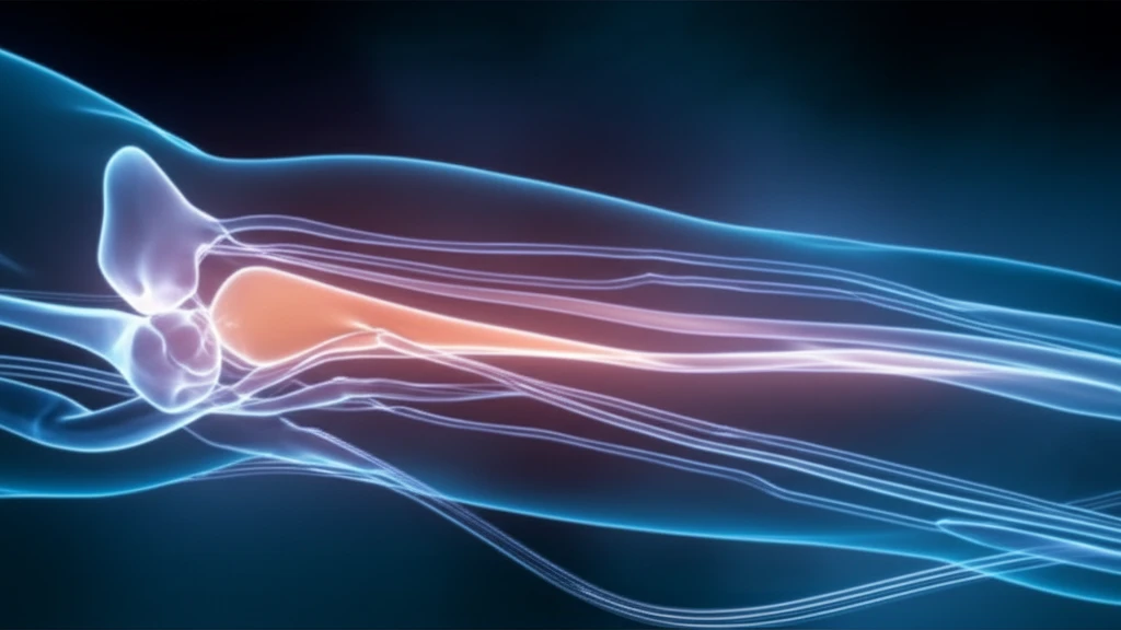
Non-contrast MRI imaging of leg arteries showing clear visualization without dye.

Non-Contrast MRI: A Game-Changer for Peripheral Artery Disease Detection?

"Discover how non-contrast MRI angiography at 3T offers comparable accuracy to traditional methods for diagnosing lower extremity arterial disease, providing a safer alternative for patients with PAD."


Peripheral artery disease (PAD) affects millions, often leading to significant mobility issues and pain. Traditionally, magnetic resonance angiography (MRI) with gadolinium-based contrast agents has been the go-to method for diagnosing PAD. However, concerns about nephrogenic systemic fibrosis and gadolinium accumulation in the brain have spurred the search for safer alternatives.

Enter non-contrast MRI angiography. Several techniques have emerged, aiming to provide accurate diagnostics without the risks associated with gadolinium. These methods capitalize on the natural properties of blood flow and advanced imaging technologies to visualize the arteries in the lower extremities.

A recent study published in the Journal of Vascular and Interventional Radiology compared two established non-gadolinium MR angiography protocols—QISS and QIR/ECG-FSE—with traditional gadolinium-enhanced MR angiography at 3T. The goal was to assess their diagnostic accuracy in detecting and characterizing lower extremity peripheral arterial disease. Here’s what they found.

Non-Contrast MRI Angiography: How Does It Work?

Non-contrast MRI imaging of leg arteries showing clear visualization without dye.

Before diving into the study's findings, it's important to understand the two non-contrast MRI techniques evaluated:

Here’s a breakdown of the two techniques used:

  • QISS (Quiescent-Interval Single-Shot): This technique uses balanced steady-state free precession readout, time-of-flight effects, and a tracking saturation band to capture images of inflowing arterial blood. It doesn't require image subtraction, making it less susceptible to motion artifacts.
  • QIR/ECG-FSE (Quadruple Inversion Recovery/Electrocardiogram-Gated Fast Spin Echo): This method combines quadruple inversion recovery for the abdominopelvic region with ECG-gated fast spin echo for the extremities. ECG gating helps to reduce motion artifacts by timing the image acquisition with the cardiac cycle.
Both techniques aim to visualize the arteries and detect any stenosis (narrowing) or occlusions (blockages) that are characteristic of PAD. The study sought to determine how well these non-contrast methods stacked up against the gold standard: gadolinium-enhanced MRI.

The Future of PAD Diagnosis: Non-Contrast MRI

The study's findings suggest that QISS and QIR/ECG-FSE MR angiography protocols offer comparable diagnostic accuracy to gadolinium-enhanced MRI, particularly in terms of specificity. This means they are good at correctly identifying patients who do not have significant arterial disease. Either protocol could serve as a viable alternative for patients with PAD, especially those who cannot receive gadolinium-based contrast agents. As technology advances and imaging times are reduced, non-contrast MRI is likely to play an increasingly important role in the diagnosis and management of PAD.

About this Article -

This article was crafted using a human-AI hybrid and collaborative approach. AI assisted our team with initial drafting, research insights, identifying key questions, and image generation. Our human editors guided topic selection, defined the angle, structured the content, ensured factual accuracy and relevance, refined the tone, and conducted thorough editing to deliver helpful, high-quality information.See our About page for more information.

Everything You Need To Know

1

What is Peripheral Artery Disease (PAD) and why is it a concern?

Peripheral Artery Disease (PAD) is a condition affecting millions, characterized by reduced blood flow to the limbs, often causing mobility issues and pain. The primary concern is that traditional diagnostic methods, like gadolinium-enhanced MRI, involve contrast agents that pose risks, especially for those with kidney problems or potential gadolinium accumulation. This has led to the exploration of safer alternatives like non-contrast MRI angiography.

2

What are the primary techniques used in non-contrast MRI angiography for PAD diagnosis?

The study highlighted two main techniques: QISS (Quiescent-Interval Single-Shot) and QIR/ECG-FSE (Quadruple Inversion Recovery/Electrocardiogram-Gated Fast Spin Echo). QISS uses balanced steady-state free precession and time-of-flight effects to visualize arterial blood flow without image subtraction, minimizing motion artifacts. QIR/ECG-FSE combines quadruple inversion recovery and ECG-gated fast spin echo to reduce motion artifacts, particularly those related to the cardiac cycle, making it a more precise method.

3

How does QISS (Quiescent-Interval Single-Shot) work in non-contrast MRI angiography?

QISS uses balanced steady-state free precession readout, time-of-flight effects, and a tracking saturation band. This combination allows capturing images of inflowing arterial blood. This method does not require image subtraction, a process that can be prone to motion artifacts, making it a more reliable technique. The goal is to visualize the arteries, identifying any narrowing (stenosis) or blockages (occlusions) indicative of PAD, without using contrast agents.

4

What are the advantages of using QIR/ECG-FSE (Quadruple Inversion Recovery/Electrocardiogram-Gated Fast Spin Echo) in non-contrast MRI angiography?

QIR/ECG-FSE offers a key advantage: it reduces motion artifacts. This technique combines quadruple inversion recovery for the abdominopelvic region with ECG-gated fast spin echo for the extremities. By synchronizing the image acquisition with the cardiac cycle via ECG gating, this method minimizes motion artifacts that could distort the images. This results in more accurate imaging of the arteries in the lower extremities, helping in the precise diagnosis of PAD.

5

What are the implications of non-contrast MRI angiography for the future of PAD diagnosis?

The study's findings suggest that both QISS and QIR/ECG-FSE provide comparable diagnostic accuracy to gadolinium-enhanced MRI, especially in terms of specificity. This means they can accurately identify patients without significant arterial disease. As a result, either protocol can serve as a safer alternative, especially for patients who cannot receive gadolinium. The future of PAD diagnosis likely involves non-contrast MRI, potentially reducing risks and improving patient care, particularly as technology advances and imaging times are reduced.

Newsletter Subscribe

Subscribe to get the latest articles and insights directly in your inbox.