
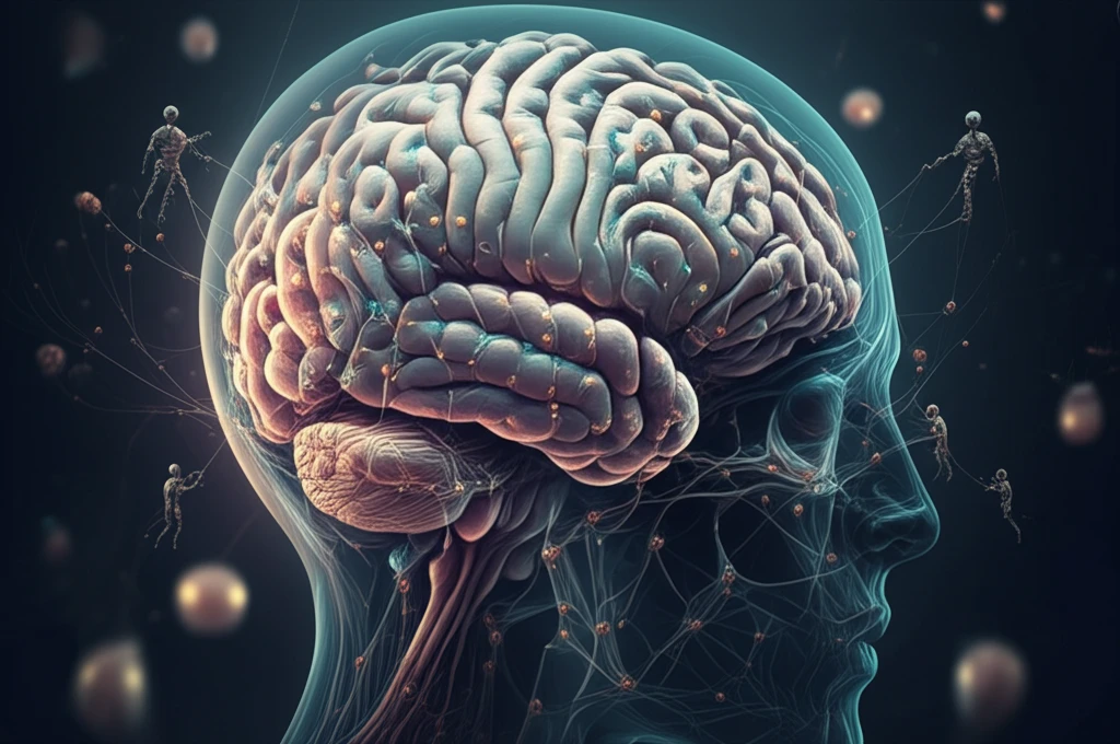
Unlock Your Brain's Potential: How Autophagy Can Protect Against Neurodegeneration

"Discover the crucial role of autophagy in maintaining brain health and how understanding its regional variations could unlock new strategies for preventing neurodegenerative diseases."
In an era where improved medical care has led to an aging population, the increasing prevalence of age-related neurological decline is a significant concern. Neurodegenerative diseases such as Alzheimer's and Parkinson's present immense social and economic challenges. While researchers have made strides in understanding the molecular mechanisms underlying these conditions, the precise causes remain elusive.
A key area of focus is why certain brain regions are more vulnerable to proteotoxicity and neuronal degradation than others. For instance, in Alzheimer's disease, the hippocampus is severely and initially affected, while the cerebellum seems relatively spared until later stages. Macroautophagy, commonly referred to as autophagy, is the main cellular process through which cells degrade misfolded proteins and impaired cytoplasmic organelles, making it vital for neuronal homeostasis and a promising target for treating neurodegenerative disorders.
Autophagy plays a dual role in neurodegeneration: it can promote degradation as a downstream effect, or disrupt proteostasis as an upstream effect, leading to protein aggregation and toxicity. Given the intricate nature of autophagy, researchers are exploring whether variations in autophagic activity across different brain regions may influence susceptibility to protein aggregation and cellular decline.
Autophagy in Disease: What the Data Shows
Autophagy is a catabolic process involved in the turnover of organelles and macromolecules that, depending on conditions, may preserve cell survival or lead to cell death. Cells launch autophagy in response to changes in internal and external environmental pressures. The relationship between autophagy-related genes and disease has received growing attention, and some lung cancer cell lines and tumor samples are characterized by increased levels of lipidated LC3, a widely used autophagy marker. Because autophagic activity varies across tissues and disease contexts, quantitative rigor matters: one gene-expression analysis used principal component analysis and z-score normalization to verify repeatability, while experimental studies commonly report means ± standard deviation with t-tests and significance set at P < 0.05. These patterns come from cancer and kidney research, so they do not provide direct statistics on neurodegeneration, but they show how autophagy is measured and linked to disease.
Measuring and Manipulating Autophagy: Tools and Limits
Standardized study of autophagy relies on shared methodology; an autophagy consortium published guidelines for standardizing autophagy research in 2008 and updated them in 2012. Because the pathway is dynamic, objective measurement is a persistent challenge, and one current approach uses multispectral imaging flow cytometry to quantify autophagy markers such as LC3 and p62 co-localization. A key limitation of pharmacological tools is that they are not autophagy-specific, as repurposed drugs inevitably have pharmacological effects beyond autophagy regulation. Interpretation is further complicated by context: inducing autophagy may be beneficial for cancer prevention, yet established tumors can use enhanced autophagy to survive metabolic and therapeutic stress. Together, these factors mean that both measuring and manipulating autophagy require carefully controlled, standardized conditions.
From Self-Eating to a Field of Its Own
The word 'autophagy' comes from 'auto,' meaning self, and 'phagy,' meaning eat, capturing the body's way of cleaning out damaged cells. It is the natural process of breaking down and removing damaged proteins and substances in the cytoplasm, unfolding through stages with distinct sets of components, and it tends to slow with age. Historical accounts trace the field's development through key milestones and document the current status of autophagy research. That history connects directly to modern questions, including whether declining autophagic activity contributes to conditions such as Alzheimer's disease and age-related brain decline.
The Brain's Clean-Up Crew: How Autophagy Works
Autophagy is essential for maintaining a healthy balance within our cells. Think of it as the brain's cellular housekeeping process, responsible for clearing out damaged or unnecessary components to keep everything running smoothly. This process involves several key steps:
- LC3-II Turnover: By blocking or inducing LC3-II degradation, researchers can monitor changes in cellular levels.
- P62 Degradation: P62, also known as SQSTM1, links LC3 and ubiquitinated substrates, facilitating their degradation via autophagy.
- Lysosomal Degradation Inhibition: Agents like chloroquine or bafilomycin A1 are used to inhibit lysosomal degradation and observe the accumulation of LC3-II and p62.
The Evolving Autophagy Research Landscape
Autophagy is a process by which cellular material is degraded by lysosomes or vacuoles and recycled. Several pathways operate within a cell, including macroautophagy, microautophagy, and chaperone-mediated autophagy, and all share the degradation of intracellular components via the lysosome. It is a self-degradative process important for balancing energy sources at critical times in development and in response to nutrient stress. Research news outlets report a steady flow of new findings, with science and medical news services tracking advances and breakthroughs in the field. This ongoing coverage reflects an active research landscape in which autophagy's roles in health and disease continue to be refined.
When Autophagy Is Not the Hero
Despite its reputation as a purely protective process, the evidence shows autophagy is not universally beneficial. A review of diabetes research found both beneficial and detrimental roles for autophagy in diabetes mellitus and its comorbidities. In critical illness, autophagy is implicated as a crucial repair process needed to recover from organ failure, but the balance can be delicate. Foundational experiments also show how readily the machinery can fail: researchers identified 15 autophagy-defective yeast mutants that failed to form autophagic bodies. These caveats complicate simple 'boost your autophagy' messaging, since outcomes depend heavily on context, disease stage, and the organism's state.
Autophagy, Apoptosis, and the Proteasome: Three Clearance Systems
Autophagy is often compared with apoptosis, another form of regulated cell death that occurs naturally in living cells and is mediated by immune responses as a defense mechanism. Unlike apoptosis, which dismantles the cell, autophagy recycles damaged content and can even help cancer cells survive, since tumors use autophagy as a way to deal with cellular stress. Autophagy also works alongside other clearance machinery, especially the ubiquitin-proteasome system; because both are critical for cellular homeostasis, covering ATP balance, amino acid recycling, and protein quality control, their activities must be carefully orchestrated. These distinctions matter for interpreting results: the same pathway that supports cellular self-maintenance in healthy tissue can, in the wrong context, sustain disease.
The Future of Brain Health: Harnessing Autophagy
The findings suggest that variability in basal autophagic activity across different brain regions may contribute to the region-specific decline observed in neurodegenerative diseases like Alzheimer's and Parkinson's. Regions with lower basal autophagic activity, such as the hippocampus, may be more vulnerable to proteotoxic stress, while regions with higher activity, like the cerebellum, may be better protected. Future research should focus on in-depth analyses of brain-region-specific autophagic flux to develop targeted therapies that can prevent or halt neurodegenerative disease progression.
What the Experts Emphasize
Expert commentary consistently emphasizes that autophagy's effects are context-dependent. One expert opinion notes that autophagy/mitophagy activation during reperfusion after ischemia contributes to brain damage, including neuronal cell death, blood-brain barrier disruption, and neurological deficits. Similarly, reviews of hepatic steatosis examine the pathophysiological role of autophagy in liver fat accumulation. Autophagy researcher Guido Kroemer highlights that several lifestyle behaviors can induce autophagy, including exercise, fasting, and dietary intake of caloric restriction mimetics. Because apoptosis and autophagy both impact cell death across multiple body systems, experts caution that new therapeutic strategies must account for their complex roles during development and in modulating toxic cellular environments.
Precision Medicine and the Double-Edged Sword
The future outlook for autophagy research centers on precision medicine and on recognizing that the pathway is a double-edged sword. In cancer, autophagy has controversial roles: during the early stage it appears protective, suppressing malignant transformation, while later it can promote tumor progression. This duality has prompted researchers to ask whether targeting autophagy in cancer is a good approach and to explore how the pathway might be exploited for precision medicine. Looking ahead, investigators describe autophagy as a degradative process that clears worn-out proteins and organelles, maintaining cellular homeostasis and genomic stability by removing toxic waste and regulating reactive oxygen species, positioning it as a candidate target in age-related disease. Popular 2026 guides increasingly translate this science into anti-aging practice, covering fasting protocols, clinical trials, benefits, risks, and long-term longevity challenges.
Autophagy Within the Larger Biological System
In the broader context of human disease, defective autophagy carries particular pathophysiological importance for neurodegeneration, and the modern definition of autophagy encompasses both non-selective and selective forms. Autophagy is a cytoprotective mechanism triggered by diverse stimuli, including nutrient deprivation, oxidative stress, hypoxia, protein aggregates, and toxic molecules, to mitigate stress. Yet the pathway does not operate in isolation: research on the interplay between reactive oxygen species and autophagy tracks its changing role across cancer development, from cellular transformation to metastasis. At a systems level, molecules such as heparan sulfate proteoglycans influence autophagy, membrane trafficking, mitochondrial quality control, and lipid metabolism, showing how autophagy is embedded within larger regulatory networks. These system-level connections underscore why simple interventions are unlikely to produce simple answers for complex diseases.
What Autophagy Means for Real People
For general audiences, the human scale of the system is striking: the body contains roughly 30 trillion cells, with about 50–70 billion created each day, and many errors occur along the way. Autophagy is indeed essential for healthy cells, and it is also true that problems with autophagy are associated with some diseases, yet as one leading health organization cautions, 'autophagy isn't that simple.' Some studies have detected autophagy in cultured human neutrophils after 24 hours, but there are no conclusive human studies identifying an optimal fasting period to achieve autophagy. Popular guides nevertheless present real-world success stories, including case studies of pathologies addressed through natural autophagy protocols involving fasting, foods, and lifestyle routines. Taken together, the evidence suggests enthusiasm for autophagy-based interventions should be balanced with realistic expectations until more definitive human data emerges.