
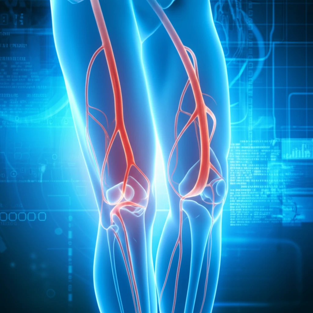
Illustration of leg arteries using non-contrast MRI

Beyond the Scan: Revolutionizing PAD Detection with Non-Contrast MR Angiography

"New Research Highlights Cutting-Edge Imaging Techniques to Diagnose Peripheral Artery Disease Without Invasive Procedures."


Peripheral Artery Disease (PAD), a condition marked by the narrowing of arteries, often goes unnoticed until it progresses, potentially leading to severe complications like limb amputation. Traditional diagnostic methods often involve invasive procedures or the use of contrast agents, which carry risks. However, recent advancements in medical imaging are offering new hope. Non-contrast MR angiography is emerging as a powerful, non-invasive tool, promising a safer and more effective way to detect and characterize PAD.

This article delves into a groundbreaking study that compares the diagnostic accuracy of non-contrast MR angiography protocols with the conventional, contrast-enhanced method. The research focuses on two primary non-contrast techniques performed at 3T (a type of MRI machine), assessing their ability to evaluate lower extremity PAD. The goal is to determine if these innovative methods can match the effectiveness of traditional approaches while minimizing patient risk.

The study's findings have significant implications for patient care, offering potential alternatives that could transform how PAD is diagnosed and managed. By exploring these new approaches, we can better understand the potential for safer, more accurate, and more accessible diagnostic tools in the fight against PAD.

AI Search Multiple angles on this topic

Evidence for Non-Contrast Imaging

Studies have evaluated non-contrast quiescent interval single-shot (QISS) magnetic resonance angiography at 3T for lower-extremity peripheral arterial disease, including comparisons with CT angiography. Research has also assessed ECG-gated QISS MRA in patients with advanced peripheral arterial occlusive disease. These investigations position non-contrast MRA as a potentially useful approach for evaluating PAD while avoiding contrast administration.

Imaging Choices and Tradeoffs

PAD assessment can draw on magnetic resonance angiography, CT angiography, duplex ultrasonography, and physiologic testing, with each modality providing different anatomic or physiologic information. Non-contrast MRA is described as an alternative to contrast-enhanced MRA or CT, but prolonged examination times and potentially inferior diagnostic performance remain limitations. It also offers a radiation-free alternative to CT angiography, although technique-specific physical and clinical limitations must be considered.

From Early Techniques to QISS

Peripheral MR angiography has developed through approaches including step-by-step, hybrid, continuous-table-movement, and non-contrast-enhanced techniques. Earlier lower-extremity non-contrast MRA relied on ECG-gated two-dimensional time-of-flight imaging, which was associated with very long acquisition times and variable image quality, particularly near disease and vessel tortuosity. Later work evaluated ECG-gated QISS MRA at 3T, while reviews published in 2019 and 2024 describe the continuing expansion of non-contrast MRA, especially for patients with renal complications, pregnant women, and children.

Unveiling the Power of Non-Contrast MR Angiography

Illustration of leg arteries using non-contrast MRI

Non-contrast MR angiography utilizes advanced imaging techniques that do not require the use of gadolinium-based contrast agents. This is a significant advantage, as contrast agents can pose risks, including allergic reactions and nephrogenic systemic fibrosis, particularly for patients with kidney problems. The core of these techniques lies in capturing detailed images of blood vessels without the need for these agents.

The research focuses on two key non-contrast MR angiography protocols: Quiescent-interval single-shot (QISS) and a combination of quadruple inversion recovery (QIR) with electrocardiogram-gated fast spin echo (ECG-FSE). QISS is a technique that captures images during a specific interval, minimizing motion artifacts and allowing for detailed visualization. QIR/ECG-FSE combines QIR, which enhances the clarity of blood vessels, with ECG-FSE, which synchronizes imaging with the heart's rhythm to reduce motion blur. These methods offer a way to achieve high-quality images without the risks associated with contrast agents.

  • QISS (Quiescent-interval single-shot): Uses a single-shot approach, minimizing motion artifacts and providing clear vessel images.
  • QIR (Quadruple Inversion Recovery): Improves vessel clarity by using a specific preparation sequence.
  • ECG-FSE (Electrocardiogram-gated fast spin echo): Synchronizes imaging with the heart's rhythm, reducing motion blur.
AI Search Multiple angles on this topic

An Evolving Evidence Base

Recent research continues to examine non-contrast MR angiography as a diagnostic approach for PAD. Reviews describe multiple bright-blood non-contrast MRA techniques and discuss their physical principles, limitations, and clinical applications. The available direction of research is promising, but the evidence should be interpreted according to the specific technique, patient population, and imaging comparison used.

Recognized Limitations

Non-contrast MRA should not be treated as free of practical limitations. Potential concerns include longer examination times, variable image quality, and diagnostic performance that may be inferior to established contrast-enhanced or CT-based approaches in some settings. These limitations suggest that implementation requires attention to patient selection, protocol quality, and local expertise.

Different Modalities, Different Strengths

No single PAD imaging method necessarily answers every clinical question. MRA, CTA, duplex ultrasonography, and physiologic testing contribute different types of anatomic and functional information, while non-contrast MRA is attractive when contrast exposure or radiation is undesirable. Meaningful comparison therefore depends on the clinical purpose, the patient's risks, and the performance of the selected protocol.

The study compared these non-contrast methods to contrast-enhanced MR angiography, the standard diagnostic approach. The research involved a detailed evaluation of patients with PAD, comparing the diagnostic accuracy of each technique. The researchers assessed the ability of each method to identify and characterize arterial abnormalities. The results provide critical insights into the effectiveness and reliability of these innovative imaging techniques, offering a pathway to improved patient care.

The Future of PAD Diagnosis: A Brighter Outlook

The research highlights the promising potential of non-contrast MR angiography in the diagnosis of PAD. As technology continues to evolve, these methods could become the new standard of care, offering a safer and more effective approach to diagnosing and managing this widespread condition. By embracing these advancements, healthcare providers can enhance patient care and improve outcomes for those affected by PAD.

AI Search Multiple angles on this topic

A Complementary Diagnostic Role

Non-contrast MR angiography is best understood as an expanding option within the broader PAD imaging toolkit. Its value is particularly apparent when clinicians seek vascular assessment without contrast agents, but its limitations mean it should be integrated thoughtfully rather than viewed as a universal replacement. Expert interpretation remains important because image quality and diagnostic performance can vary across techniques and clinical circumstances.

Toward Safer Quantitative Assessment

Future work is exploring non-contrast MRA as a safer alternative for PAD detection, including 3T imaging for people with kidney concerns. A 2025 study of three-station lower-extremity NC-MRA describes the technique as a safe and quantitative approach for early PAD detection without contrast-agent risks. Reviews also note that MRA can delineate vascular patency without the dense mural-calcification problem that can affect CT angiography.

Implementation Beyond the Image

Wider adoption of non-contrast MRA may depend on more than technical capability alone. Health systems may need appropriate equipment, trained personnel, standardized protocols, and workflows that accommodate examination-time demands. Because these practical factors can differ across institutions, the technology's real-world impact may develop unevenly.

Balancing Safety and Convenience

For people with lower-extremity PAD, non-contrast MRA offers a potential alternative to contrast-enhanced MRA or CT. The cited review also cautions that it may involve prolonged examinations and inferior diagnostic performance, so the patient benefit is not simply a matter of removing contrast. Clinical decisions must balance safety considerations with scan duration, image quality, and the diagnostic needs of the individual.

QISS accuracy against contrast-enhanced MRA

  • 94.1% sensitivity; 97.8% specificityIn patients with advanced peripheral arterial occlusive disease, HR-QISS detected significant stenosis with 94.1% sensitivity and 97.8% specificity, using contrast-enhanced MRA as the reference.[1]

What accuracy figures show-and what they do not

The high accuracy reported in a small subset is encouraging, but its small size makes it an uncertain guide to performance across a broader PAD population.[2]

Including people with good vascular status may make QISS appear more accurate than it would be in more challenging cases of arterial disease.[1]

Later work describes 3D ECG-FSE as improving the display of slow-flowing blood and collateral arteries while reducing motion artifacts; another account characterizes it as enabling lower-artery visualization without motion artifacts. These are reported technical capabilities, not direct accuracy comparisons with QISS.[3]

What remains unresolved for non-contrast MRA

  • How well do accuracy estimates from advanced-disease cohorts transfer to people with less severe disease, given the concern that including people with good vascular status can inflate estimates?[1]
  • How much does the choice of reference standard affect apparent QISS performance, when reported comparisons use contrast-enhanced MRA, digital subtraction angiography, or both?[1] [2] [4]
  • Can the reported imaging benefits of 3D ECG-FSE, including visualization of slow flow and collateral arteries, be translated into consistently better diagnostic decisions?[3]

Terms used in the evidence

Flow-sensitive dephasing
A balanced steady-state approach[5]

About this Article -

Written with AI assistance from published research, and reviewed by the Mystum team. See our About page for more information.

This article is based on research published under:

DOI-LINK: 10.1016/j.jvir.2018.06.015, Alternate LINK

Title: Diagnostic Accuracy Of Noncontrast Mr Angiography Protocols At 3T For The Detection And Characterization Of Lower Extremity Peripheral Arterial Disease

Subject: Cardiology and Cardiovascular Medicine

Journal: Journal of Vascular and Interventional Radiology

Publisher: Elsevier BV

Authors: Christopher J. Hanrahan, Marc D. Lindley, Michelle Mueller, Daniel Kim, Daniel Sommers, Glen Morrell, Andrew Redd, Kristi Carlston, Vivian S. Lee

Published: 2018-11-01

Everything You Need To Know

1

What is Peripheral Artery Disease (PAD), and why is early detection so important?

Peripheral Artery Disease (PAD) is a condition characterized by the narrowing of arteries, most commonly in the legs. Early detection is crucial because PAD often goes unnoticed until it progresses significantly, potentially leading to severe complications, including limb amputation. Identifying and managing PAD early can help prevent these adverse outcomes through lifestyle changes, medication, or interventions to improve blood flow. Non-contrast MR angiography offers a safer way to detect PAD without invasive procedures or contrast agents, aiding in earlier and more accurate diagnoses.

2

How does non-contrast MR angiography differ from traditional methods for diagnosing PAD, and what are its advantages?

Traditional methods for diagnosing Peripheral Artery Disease (PAD) often involve invasive procedures or the use of contrast agents, which can carry risks such as allergic reactions or nephrogenic systemic fibrosis, especially for patients with kidney problems. Non-contrast MR angiography, on the other hand, uses advanced imaging techniques that do not require gadolinium-based contrast agents. This approach reduces patient risk while still providing detailed images of blood vessels, making it a safer and more effective alternative for detecting and characterizing PAD.

3

Could you explain the specific non-contrast MR angiography techniques, such as QISS and QIR/ECG-FSE, and how they work to visualize blood vessels?

The research highlights two primary non-contrast MR angiography protocols: Quiescent-interval single-shot (QISS) and a combination of quadruple inversion recovery (QIR) with electrocardiogram-gated fast spin echo (ECG-FSE). QISS captures images during a specific interval, minimizing motion artifacts for detailed visualization. QIR enhances the clarity of blood vessels, while ECG-FSE synchronizes imaging with the heart's rhythm to reduce motion blur. Together, these methods achieve high-quality images of blood vessels without needing contrast agents, providing a safer and effective way to diagnose Peripheral Artery Disease (PAD).

4

What are the implications of using non-contrast MR angiography for patients with kidney problems, and how does it improve their safety during PAD diagnosis?

Patients with kidney problems are at higher risk when undergoing traditional contrast-enhanced imaging due to the potential for nephrogenic systemic fibrosis. Non-contrast MR angiography eliminates this risk by not requiring gadolinium-based contrast agents. By using techniques like Quiescent-interval single-shot (QISS) and a combination of quadruple inversion recovery (QIR) with electrocardiogram-gated fast spin echo (ECG-FSE), healthcare providers can safely and effectively diagnose Peripheral Artery Disease (PAD) in these vulnerable patients, avoiding complications associated with contrast agents and improving their overall care.

5

If non-contrast MR angiography becomes the standard for PAD diagnosis, how might this impact the accessibility and cost-effectiveness of screening for Peripheral Artery Disease?

If non-contrast MR angiography becomes the standard for diagnosing Peripheral Artery Disease (PAD), it could significantly enhance accessibility and potentially improve cost-effectiveness. By eliminating the need for contrast agents, it reduces the risk of adverse reactions and the associated costs of managing those reactions. Techniques such as Quiescent-interval single-shot (QISS) and quadruple inversion recovery (QIR) with electrocardiogram-gated fast spin echo (ECG-FSE) can be implemented more widely, particularly in settings where contrast-enhanced imaging is limited or contraindicated. This shift could lead to earlier and more widespread screening, ultimately improving patient outcomes and reducing the overall burden of PAD.

Newsletter Subscribe

Subscribe to get the latest articles and insights directly in your inbox.