
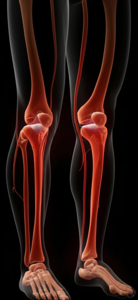
Non-Contrast MRI: A Safer Way to Detect Peripheral Artery Disease?

"Discover how non-contrast MRI angiography offers a radiation-free and gadolinium-free alternative for diagnosing PAD, making it safer for patients with kidney concerns."
Peripheral artery disease (PAD) affects millions, often leading to pain and reduced mobility. Traditional diagnosis relies on methods like contrast-enhanced magnetic resonance angiography (MRA), which uses gadolinium-based contrast agents. However, these agents pose risks, especially for individuals with kidney problems.
Gadolinium-based contrast agents have been linked to nephrogenic systemic fibrosis and gadolinium accumulation in the brain, prompting the search for safer alternatives. Non-contrast MRA techniques are emerging as promising options, offering a way to visualize blood vessels without contrast agents.
This article explores the accuracy of non-contrast MRA protocols at 3T (Tesla) for detecting and characterizing lower extremity PAD. We'll delve into how these techniques compare to gadolinium-enhanced MRA, providing insights into their effectiveness and potential benefits for patients.
Accepted Imaging Methods
Contrast-enhanced magnetic resonance angiography provides excellent assessment of the peripheral arterial vasculature and is an important adjunct for evaluating peripheral artery disease. However, the high prevalence of chronic kidney disease among patients with peripheral artery disease raises concerns about gadolinium contrast media. MRA can also provide high-quality arterial imaging for patients previously considered unsuitable for contrast studies and is minimally affected by arterial calcification, supporting luminal evaluation in diabetic or renal patients with dense medial sclerosis. Noncontrast MRA offers a safe, quantitative approach for early PAD detection without contrast-agent risks.
From MRA to QISS
Magnetic resonance angiography emerged as a standard noninvasive imaging method for peripheral artery disease, with diagnostic accuracy compared with percutaneous angiography gold standards. The development of TRANCE-MRI marked a milestone by enabling noninvasive imaging of arteries and veins in the lower extremities without contrast. Later work on noncontrast techniques included QISS MRA, illustrated by a case involving a 67-year-old man with diabetes, hypercholesterolemia, hypertension, worsening PAD symptoms, and a prior bypass from the left common femoral artery to the posterior tibial artery. These developments helped focus MRI research on patients who may face risks from gadolinium-based contrast agents.
Understanding Non-Contrast MRI Angiography Techniques
Non-contrast MRA utilizes various techniques to visualize blood vessels without contrast agents. These methods rely on the natural properties of blood flow and magnetic fields to create images of the arteries. Two established non-contrast MRA protocols are quiescent-interval single-shot (QISS) and a combination of quadruple inversion recovery (QIR) with electrocardiogram-gated fast spin echo (ECG-FSE).
- Quiescent-Interval Single-Shot (QISS): This technique employs balanced steady-state free precession readout, time-of-flight effects, and a tracking saturation band to image inflowing arterial blood. QISS does not require image subtraction, making it less sensitive to motion artifacts.
- Quadruple Inversion Recovery (QIR) with ECG-Gated Fast Spin Echo (ECG-FSE): QIR is used for the abdominopelvic station, while ECG-FSE is used for the extremities. ECG-FSE relies on signal intensity differences between fast-flowing arterial blood during systole and slow-flowing blood during diastole. Images of arterial blood are obtained by subtraction, eliminating signal from venous blood and stationary tissues.
New Directions in NC-MRA
Recent research has examined noncontrast quiescent-interval slice-selective MRA combined with MRI-based visualization of vascular calcification for assessing arterial stenosis in patients with lower-extremity peripheral artery disease. A 2024 review describes advances in noncontrast MRA and its growing importance in body imaging. The review particularly highlights potential relevance for patients with renal complications, pregnant women, and children. Together, these studies show continuing efforts to expand the clinical role of NC-MRA while addressing patient groups for whom contrast may be undesirable.
Remaining Technical Limits
Noncontrast MRI protocols still have important limitations. One study reported that its cohort was relatively small and that multicenter research may be needed to confirm diagnostic performance across broader patient groups, indications, and clinical scenarios. Another study found only moderate inter-rater agreement for diagnostic confidence and stenosis assessment despite a training session before independent image analysis. In addition, slow flow or flow reversal caused by occlusion can challenge non-gadolinium techniques because they rely on blood flow to generate arterial signal.
QISS Versus FSE
At 3 tesla, a comparison of 2D QISS-MRA and 3D fast spin-echo MRA found significantly better diagnostic performance for QISS-MRA. The reported sensitivity was 85% versus 54%, specificity was 90% versus 47%, and diagnostic accuracy was 89% versus 48%, with p ≤ 0.0001. Separate research evaluated whether 3D prototype thin-slab stack-of-stars QISS could provide clinical benefits from superior spatial resolution compared with standard 2D-QISS, using computed tomography angiography as the reference. These findings support comparing specific noncontrast protocols rather than treating NC-MRA as a single uniform technique.
The Future of PAD Diagnosis
Non-contrast MRA techniques like QISS and QIR/ECG-FSE offer comparable diagnostic accuracies with high specificity for detecting PAD. Either protocol provides a valuable alternative to gadolinium-enhanced MRA, particularly for patients with kidney concerns or those seeking a radiation-free imaging option. As MRI technology advances, non-contrast methods are poised to play an increasingly important role in vascular imaging.
Context Matters in Interpretation
Evidence on peripheral MRA includes protocols that combine noncontrast calf imaging with large-field-of-view contrast-enhanced MRA. In one series, 16 consecutive patients with peripheral artery disease underwent a combined protocol, and digital subtraction angiography was performed within one week of MR angiography. Image quality and stenosis were assessed by two experienced readers. Related work on 3D thin-slab stack-of-stars QISS evaluated its potential clinical benefit over standard 2D-QISS using computed tomography angiography as a reference, underscoring the importance of protocol design, reader expertise, and the chosen reference test when interpreting results.