Are Intraoral Scanners the Future of Dentistry? A Deep Dive into Accuracy
"Explore how digital models created with intraoral scanners stack up against traditional methods and what it means for patient care."
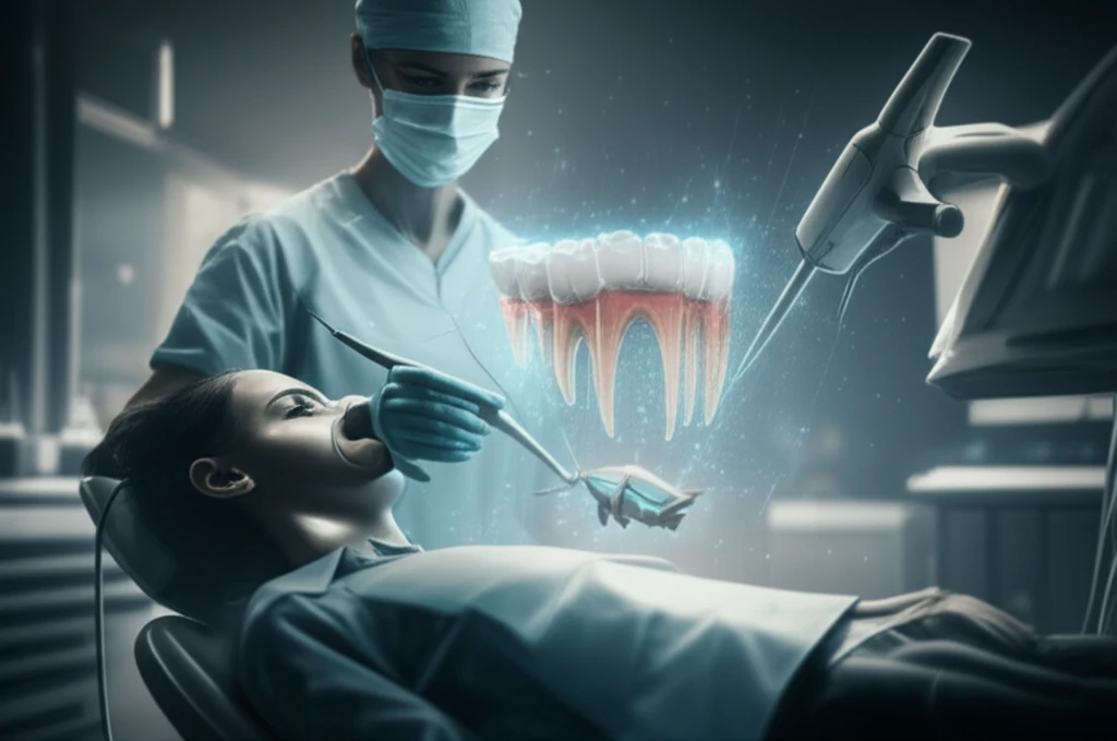
The world of dentistry is rapidly evolving, driven by advancements in technology that promise greater precision, efficiency, and patient comfort. One of the most transformative innovations is the use of digital dental models, crafted directly from intraoral scanners. These scanners eliminate the need for traditional impressions, streamlining workflows and opening new possibilities for appliance fabrication and treatment planning.
Digital models offer several advantages, including reduced chair time, minimized patient discomfort, and the potential for more precise indirect bracket setups. By incorporating digital technology, dental practices can minimize the need for bracket repositioning, ultimately decreasing overall treatment time. The appeal is clear: a faster, more comfortable experience for patients and a more efficient practice for clinicians.
However, the universal adoption of intraoral scanners has been gradual. With various methods available to digitize dental models, dental professionals need to know how accurate these newer approaches are. This article explores the accuracy of 3D measurements taken from digital models created by intraoral scanners, comparing them to manual measurements. It provides a comprehensive look into whether these scanners are truly as accurate as traditional methods.
How Accurate Are Intraoral Scanners Compared to Traditional Methods?
A recent study published in the American Journal of Orthodontics and Dentofacial Orthopedics sought to evaluate and compare digital dental models generated from two commercial intraoral scanners against manual measurements. The focus was on performing 3-dimensional surface measurements along curved lines—a critical aspect of dental morphology. The study used dry mandibles (n = 61) with intact dentition, digitized using the Cadent iTero and Lythos Digital Impression System. These models were then converted into stereolithography files for measurement using specialized software. Manual measurements were taken directly on the mandibular teeth for comparison.
- No Significant Differences: The study found no significant differences between any of the paired methods (P > 0.05), indicating a level of agreement between the intraoral scanners and manual measurements.
- Random Errors Detected: Bland-Altman analysis revealed no fixed bias in any approach, but random errors were detected across all comparisons.
- Low Mean Bias: The mean biases of digital models obtained by iTero and Lythos scanners were low when compared to direct caliper measurements. The comparison between the two intraoral scanners yielded the lowest mean bias.
- No Proportional Bias: No comparison displayed statistical significance for t-scores, suggesting an absence of proportional bias.
The Future of Digital Dentistry
The study's conclusion highlights that intraoral scanners can produce digital dental models that are comparatively accurate for direct surface measurements along curved lines in 3D. As technology continues to advance, the integration of intraoral scanners into dental practices promises to enhance efficiency, precision, and patient experience. Although challenges remain, the evidence suggests that intraoral scanners are poised to play a pivotal role in the future of digital dentistry, providing clinicians with reliable and accurate tools for diagnosis, treatment planning, and appliance fabrication.