Endoscopic view of a spine with highlighted pain generators

Unlocking Back Pain Relief: How Endoscopic Spine Surgery Visualizes and Treats Pain Generators

"Discover the innovative approach of in-vivo endoscopic visualization for diagnosing and surgically treating pain at its source, offering new hope for chronic back pain sufferers."


For years, managing back pain has largely relied on methods that only offer short-term fixes. Traditional approaches, such as injections and medication, often mask the symptoms without addressing the root cause. These treatments depend on the body's own healing processes to reduce pain, which can be unpredictable and inconsistent.

However, a groundbreaking approach is changing the landscape of back pain treatment. By using an endoscope to visualize the specific areas causing pain, surgeons can now target and treat these pain generators directly. This method opens the door for surgical decompression and ablation, precisely addressing the source of the pain.

Endoscopic spine surgery stands out due to its ability to use specialized tools and techniques, including mobile cannulas, advanced endoscopes, and RF and laser technology, to effectively treat the pain generator. While traditional surgeries involve large incisions and more extensive tissue disruption, endoscopic methods offer a less invasive alternative, especially when combined with intradiscal therapies.

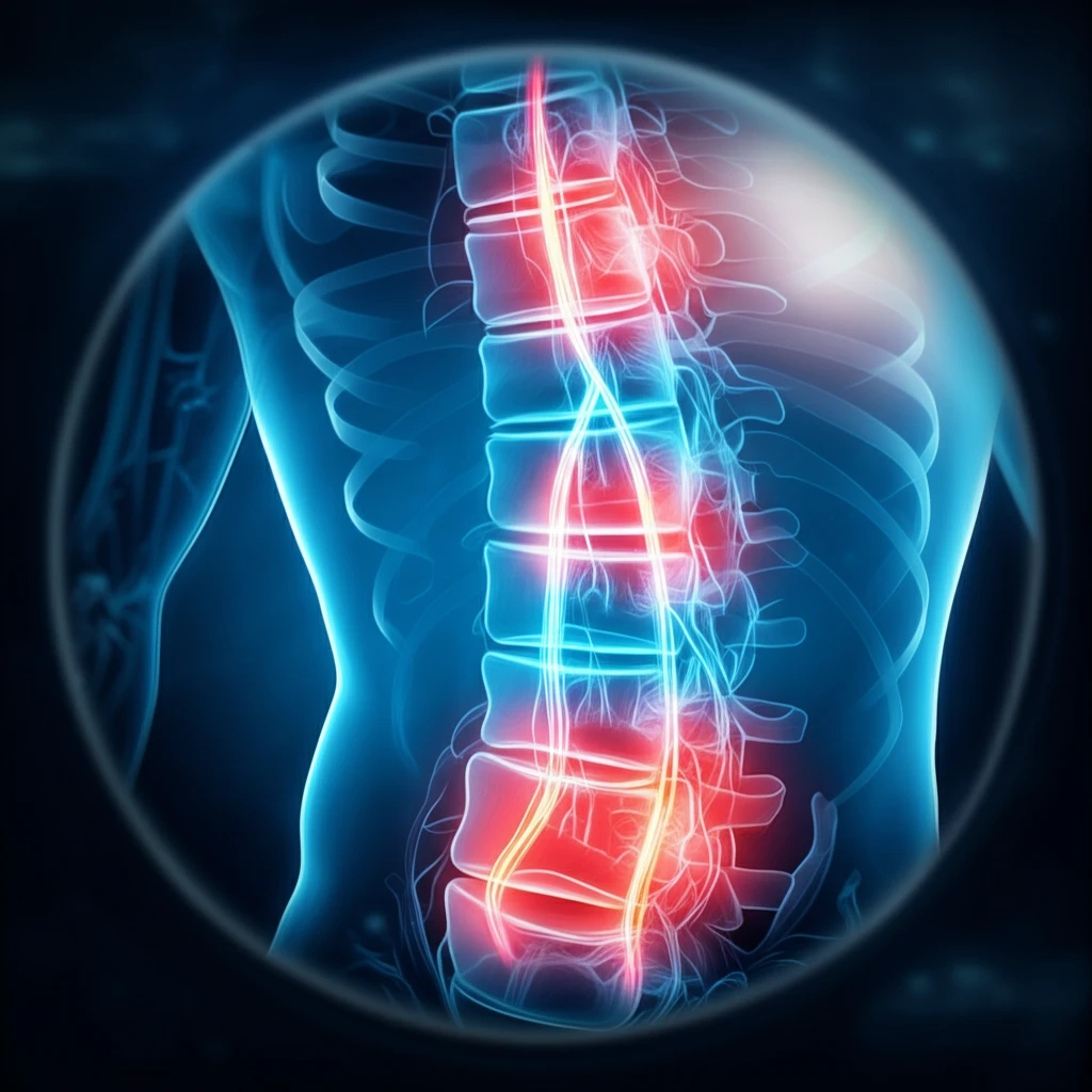
Visualizing the Invisible: How Endoscopy Pinpoints Pain Sources

Endoscopic view of a spine with highlighted pain generators

The Yeung Endoscopic Spine Surgery™ (YESSTM) technique represents a significant advancement in how we approach spinal pain. This method uses a series of precise steps to identify and treat pain generators within the spine:

The procedure begins with careful planning using X-ray imaging to determine the optimal placement for needles and cannulas. This ensures accurate access to the targeted area. The path of the needle is similar to that used for diagnostic and therapeutic injections, helping to predict the success of the endoscopic procedure.

  • Epiduralgram: A non-ionic contrast is injected to create a foraminal epiduralgram, which highlights the anatomy of the area and reveals potential issues like herniated discs or stenosis.
  • Chromo-Discography: This involves injecting dye into the disc to assess its integrity and identify any tears or abnormalities that could be causing pain.
  • Tissue Staining: Vital tissue staining helps differentiate between healthy and unhealthy tissue, guiding the surgeon during decompression.
  • Foraminoplasty: The lateral recess is decompressed, and the exiting and traversing nerves, along with the dorsal root ganglion (DRG), are carefully visualized. The endoscope allows the surgeon to see anomalies that might be missed by traditional imaging.
  • Exploration: The epidural space is thoroughly examined, and the “hidden zone” of Mac Nab is probed under local anesthesia, allowing the patient to provide feedback during the procedure.
  • Removal: Extruded or sequestered nucleus pulposus and other problematic tissues are removed using biportal or multiple portal techniques.
  • Rhizotomy: The branches of the dorsal ramus are carefully ablated to denervate the facet joint, reducing pain signals.
By documenting over 10,000 surgical cases with detailed video imaging, surgeons can identify which painful conditions are most suitable for this endoscopic approach.

The Future of Spine Care: Minimally Invasive and Highly Visual

Endoscopic spine surgery requires specialized skills and training for spine surgeons. By incorporating interventional pain management techniques with surgical precision, and by confirming the results under local anesthesia, we can ensure rational and effective treatment of spinal pain. New procedures focusing on intradiscal therapy, disc augmentation, biologics, annular modulation, and tissue neuromodulation are well-suited for this minimally invasive approach. As technology evolves with robotics and biologics, endoscopic foraminal access to the lumbar spine promises to transform spine care, offering true minimally invasive access without destabilizing the dorsal muscle column.

About this Article -

This article was crafted using a human-AI hybrid and collaborative approach. AI assisted our team with initial drafting, research insights, identifying key questions, and image generation. Our human editors guided topic selection, defined the angle, structured the content, ensured factual accuracy and relevance, refined the tone, and conducted thorough editing to deliver helpful, high-quality information.See our About page for more information.

This article is based on research published under:

DOI-LINK: 10.4172/2165-7939.1000385, Alternate LINK

Title: In-Vivo Endoscopic Visualization Of Pain Generators In The Lumbar Spine

Subject: Industrial and Manufacturing Engineering

Journal: Journal of Spine

Publisher: OMICS Publishing Group

Authors: Anthony T Yeung

Published: 2017-01-01

Everything You Need To Know

1

What makes Endoscopic Spine Surgery different from traditional back pain treatments?

Endoscopic spine surgery differs significantly from traditional methods because it directly visualizes and targets the pain generators. Unlike injections and medication that merely mask symptoms, endoscopic surgery uses an endoscope to pinpoint the exact source of pain, allowing for surgical decompression and ablation. This targeted approach aims for lasting relief by addressing the root cause of the pain, rather than providing temporary fixes. This is enhanced with specialized instruments like mobile cannulas and technologies such as RF and laser.

2

Can you explain the Yeung Endoscopic Spine Surgery™ (YESSTM) technique and its key steps?

The Yeung Endoscopic Spine Surgery™ (YESSTM) technique is a precise method for identifying and treating spinal pain. It involves several key steps, starting with careful planning using X-ray imaging to guide the placement of needles and cannulas. An epiduralgram uses non-ionic contrast to highlight the anatomy and reveal issues like herniated discs or stenosis. Chromo-discography assesses disc integrity by injecting dye. Tissue staining helps differentiate between healthy and unhealthy tissue. Foraminoplasty decompresses the lateral recess, visualizing nerves and the dorsal root ganglion (DRG). The epidural space is explored, and problematic tissues are removed, potentially with biportal or multiple portal techniques. Finally, rhizotomy ablates branches of the dorsal ramus to denervate the facet joint, reducing pain signals.

3

How does Endoscopic Spine Surgery visualize pain sources that traditional imaging might miss?

Endoscopic spine surgery visualizes pain sources through direct in-vivo visualization using an endoscope. During foraminoplasty, the endoscope allows the surgeon to directly see anomalies of the exiting and traversing nerves, along with the dorsal root ganglion (DRG), which might be missed by traditional imaging methods like X-rays or MRIs. The exploration phase includes probing the “hidden zone” of Mac Nab under local anesthesia, allowing patient feedback to guide the surgeon in identifying pain generators that would otherwise remain invisible. Vital tissue staining is also used to differentiate healthy from unhealthy tissue.

4

What are the potential future advancements in spine care that could further enhance Endoscopic Spine Surgery?

The future of spine care includes advancements like robotics and biologics that could enhance endoscopic foraminal access to the lumbar spine. New procedures focusing on intradiscal therapy, disc augmentation, biologics, annular modulation, and tissue neuromodulation are well-suited for this minimally invasive approach. The combination of interventional pain management techniques with surgical precision, along with confirmation under local anesthesia, promises more rational and effective treatment of spinal pain. These advancements aim to provide true minimally invasive access without destabilizing the dorsal muscle column.

5

What role do interventional pain management techniques play in Endoscopic Spine Surgery, and why is local anesthesia important?

Interventional pain management techniques are integrated into Endoscopic Spine Surgery to precisely target and treat pain generators. Techniques like epiduralgram and chromo-discography help identify the specific areas causing pain. Local anesthesia is crucial because it allows for real-time feedback from the patient during the procedure, particularly when probing the “hidden zone” of Mac Nab. This feedback helps the surgeon accurately locate and address the pain source, ensuring a more effective and rational treatment of spinal pain. This immediate feedback mechanism is not possible under general anesthesia, making local anesthesia a key component of the procedure's success.

Newsletter Subscribe

Subscribe to get the latest articles and insights directly in your inbox.