
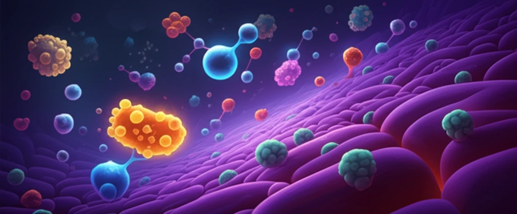
Unlock Your Cells' Secrets: How Diffusion Coefficients Reveal the Truth About Cell Membranes

"Fluorescence correlation spectroscopy offers a non-invasive peek into the dynamic world of lipids and proteins in living cell membranes, promising breakthroughs in understanding cell function."
Our cells are like bustling cities, with constant movement and interaction happening at the molecular level. The cell membrane, a flexible barrier surrounding each cell, plays a crucial role in these activities. It's not just a static enclosure; it's a dynamic interface where proteins and lipids—the building blocks of the membrane—are constantly moving and interacting.
Understanding the fluidity of the cell membrane and how its components move is vital for comprehending various cellular processes. These include cell signaling (how cells communicate), transmembrane signal transduction (how signals are transmitted across the membrane), and cell adhesion (how cells stick to each other and their surroundings). Think of it as understanding the traffic patterns within a city to grasp how the city functions.
Scientists have developed techniques to observe and measure these molecular movements. Among these, fluorescence correlation spectroscopy (FCS) stands out as a powerful tool. FCS allows researchers to detect the rapid dynamic behaviors of proteins and lipids in living cells, offering insights into the cell's inner workings without disrupting its natural state.
Reading Molecular Motion from Fluorescence Fluctuations
FCS measures the dynamics of fluorescent molecules in cells by tracking fluctuations in the number of molecules within a small volume illuminated by a thin beam of excitation light. Because the signal is inherently statistical, detailed mathematical analysis is central to the technique, and researchers note that the suitability of the models used to extract kinetic information from correlated data is a significant issue. Extensions such as lifetime-weighted FCS add fluorescence-lifetime information, enabling observation of fluctuations in the nano- to millisecond time region. Together these tools allow diffusion times and molecular brightness to be recovered from a single fluctuating signal.
The Classic FCS Workflow and Its Speed Problem
The classic FCS method, developed in the 1970s, records fluorescence intensity fluctuations and analyzes them to recover diffusion and interaction parameters, with related analyses such as photon counting histogram (PCH) using the same fluctuations while focusing on different properties of the signal. The approach has been commercialized, and researchers have applied it to tubulin oligomer formation and tubulin-drug interactions using instruments such as the Zeiss LSM 510-ConfoCor II. Dual-color fluorescence cross-correlation spectroscopy extends the method to interactions by cross-correlating signals from differently colored labels calibrated with freely diffusing standards. The technique's major drawback is practical: collecting reliable data requires long measurement times that can last for hours.
From Fluorescent Antibodies to Molecular-Level Dynamics
The roots of FCS trace back to early fluorescence-based biology: during a 1961 seminar, Albert Coons noted that "the hour of fluorescent antibody" had started, and some histories credit this fluorescent-antibody technology as the origin of what became FCS, most commonly applied to protein-protein interactions. The technique itself works by exciting molecules within a focused, stationary laser beam and analyzing the resulting fluorescence fluctuations to study dynamic processes on the molecular level. Because of its exceptional sensitivity to molecular diffusion properties, FCS has long been proposed as a valuable method for sizing small, freely diffusible particles with superior precision. Fluorescence fluctuation imaging with high time resolution is likewise regarded as holding huge potential for cellular biology and biomedicine.
What is Fluorescence Correlation Spectroscopy (FCS)?
Imagine throwing a pebble into a pond and watching the ripples spread. FCS is a bit like that, but instead of pebbles and water, it uses fluorescent molecules and a laser beam. In FCS, scientists shine a focused laser beam onto a small area of the cell membrane. They then observe the fluctuations in fluorescence intensity caused by the movement of fluorescently labeled molecules (like lipids or proteins) as they diffuse in and out of the observation area.
- High sensitivity: Detects even small changes in molecular movement.
- Non-invasive: Allows measurements in living cells without disrupting them.
- High resolution: Provides detailed information about molecular dynamics.
- Versatile: Can be used to study various molecules and processes in the cell membrane.
Pushing FCS Toward the Proteome and Living Tissues
Because it offers high-resolution spatial and temporal analysis of low-concentration biomolecules with comparable noninvasiveness, FCS remains an active research area. Recent work has demonstrated high-throughput fluorescence correlation spectroscopy capable of analyzing proteome dynamics in living cells. Advances also include tagging wild-type and oncogenic mutant alleles with different fluorescent proteins, allowing both variants to be analyzed in the same cell. On the translational side, researchers are applying single-photon FCS to measure particle concentration and mobility in living tissue for insights into drug delivery and pharmacokinetics.
When the Signal Lets You Down
Despite its strengths, FCS faces well-documented limitations. In two-photon FCS, the relatively low fluorescence count rate per molecule is the most crucial parameter determining signal-to-noise ratio and restricts the technique's applicability. Studies of diffusing quantum dots have also found that ordinary interpretations of correlation measurements are inadequate because nanocrystals naturally exhibit optical saturation. These constraints have motivated ongoing refinements, including dual-color FCCS, surveyed in comprehensive recent reviews of the field.
FCS Against Its Nearest Rivals
Direct comparisons between FCS and other fluorescence approaches help define where each technique excels. In one study, researchers compared FCS with time-resolved fluorescence anisotropy using a fluorescent dextran conjugate, illustrating how the two methods probe complementary aspects of molecular motion. A related development, near-field fluorescence correlation spectroscopy, has been proposed as an approach for studying living cell membrane dynamics. Such comparisons clarify when diffusion coefficients measured by FCS are the right choice and when alternative techniques add value.
The Future of Cell Membrane Research
Fluorescence correlation spectroscopy is a valuable tool for unraveling the complexities of the cell membrane. By measuring the diffusion coefficients of lipids and proteins, researchers can gain a deeper understanding of cell membrane fluidity, cell signaling, and other important cellular processes. This knowledge can pave the way for new treatments for diseases related to cell membrane dysfunction.
The Most Sensitive Window on the Cell Surface
Experts regard fluorescence correlation spectroscopy as capable of measuring diffusion on the cell surface with unparalleled sensitivity. In appropriate situations, researchers report that FCS can be the most sensitive and accurate method available for measuring receptor interaction and oligomerization. This makes it a uniquely powerful tool for extracting diffusion coefficients that reveal the behavior of receptors directly within the membrane environment.
From Lifetime Tags to Spectral Dimensions
Looking ahead, researchers are expanding FCS along several fronts. Fluorescence lifetime correlation spectroscopy (FLCS) adds lifetime-based discrimination, and the concept of fluorescence spectral correlation spectroscopy is being outlined as a further extension. Statistical rigor continues to matter, with published analyses examining the standard deviation and bias of FCS measurements. These developments build on the technique's origin in the early 1970s, when it was introduced to measure the kinetics of chemical reactions under zero-perturbation conditions.
FCS Alongside Photobleaching and Beyond
Within the wider ecosystem of biophysical measurement, FCS has long been combined with other fluctuation-based methods; as early as 1983, studies addressed fluorescence correlation spectroscopy and photobleaching recovery for multiple binding reactions. Newer extensions such as two-dimensional FCS dissect component spectra using the time correlation function, collecting fluorescence intensity at various wavelengths upon an external perturbation. This illustrates how FCS concepts continue to migrate into hybrid techniques that address increasingly complex biological systems.
Single Molecules, Supramolecular Answers
Researchers present FCS as a powerful spectroscopic tool for studying supramolecular dynamics with single-molecule sensitivity. Its practical reach is expanding, with realizations such as four-color fluorescence correlation spectroscopy achieved in a grating-based detection platform. For investigators seeking to understand cell membranes, this translates into an ability to observe individual molecular events and convert them into quantitative diffusion coefficients.