
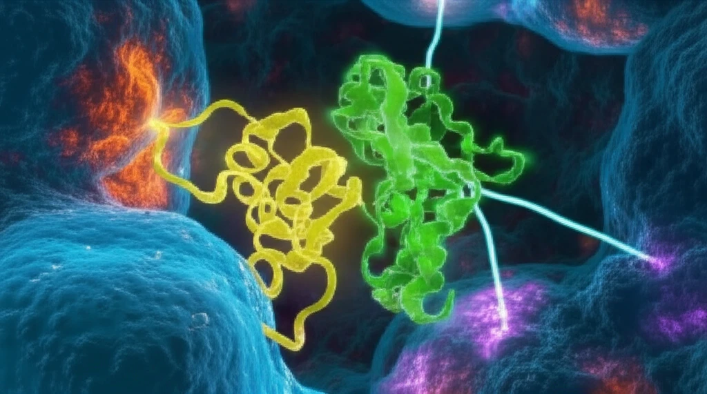
Decoding Protein Interactions: How Flexibility and Dynamics Shape Cellular Processes

"Unlocking the Secrets of Protein Binding: A Look into Intrinsically Disordered Proteins and Their Role in Cellular Signaling"
Proteins are the workhorses of our cells, and their interactions dictate nearly every biological process. Understanding how proteins bind and interact with each other is essential for unraveling the complexities of cellular function and developing new therapeutic strategies.
Intrinsically disordered proteins (IDPs) are a unique class of proteins that lack a fixed three-dimensional structure. Instead, they exist as dynamic ensembles of conformations, allowing them to interact with multiple partners and participate in a wide range of cellular processes. Their flexibility makes them difficult to study, but also crucial for cellular regulation.
This article delves into recent research exploring the conformational changes and binding mechanisms of IDPs, particularly focusing on their interactions with other proteins and the implications for cell signaling. We'll explore how techniques like molecular dynamics simulations are helping scientists understand these dynamic interactions and how they impact cellular processes.
A Vast, Shape-Shifting Proportion of the Proteome
Intrinsically disordered proteins (IDPs) lack a fixed tertiary structure, yet characterization efforts — such as the IDEAL database ("Intrinsically Disordered proteins with Extensive Annotations and Literature") and its labeled dataset of 90,253 unique protein–protein pairs drawn from DisProt — have shown these molecules are anything but inert. Advances in characterizing IDPs continue to unveil a remarkably complex interaction landscape, including coupled folding and binding, highly dynamic complexes, multivalent interactions, and even interactions between entirely disordered proteins. Because IDPs are widespread in eukaryotes and use their conformational dynamics to support a large repertoire of intra- and intermolecular interactions, they anchor a broad range of cellular processes. A 2026 functional map of human intrinsically disordered regions, built by mapping disorder features across thousands of human IDRs, further suggests which conserved aspects of disorder contribute to specific functions and interaction networks and which are associated with disease-risk genes, charting what the authors call the hidden logic of information encoded in disorder.
Measuring the Unforeseen: Methods and Their Limits
Studying proteins that lack a stable structure has required researchers to adapt techniques originally built for folded proteins. In practice, approaches typically combine solution-based biophysical measurements with computational simulations to infer the ensembles a disordered protein samples, alongside predictions from disorder-prediction algorithms. Each method carries limitations: experimental probes tend to average over many conformations, while computational models depend heavily on the training data and force fields used. Accordingly, reported disorder assignments and interaction behaviors can vary across studies, and individual findings should be read with those caveats in mind.
From Curiosities to a Structural Frontier
For much of the field's early history, the assumption that a protein's function requires a well-defined three-dimensional structure dominated biology, leaving unstructured proteins to be treated as curiosities or experimental artifacts. Observations that some biologically active proteins resisted folding gradually accumulated, and the formal concept of intrinsic disorder emerged out of these anomalous findings. The development of dedicated databases and prediction tools marked the shift from isolated examples to a recognition that disorder is common, particularly across eukaryotic proteomes. As these ideas spread, what once seemed a contradiction of the structure–function paradigm became a research field in its own right, though the precise chronology and attribution of early milestones differ somewhat across historical accounts.
The Dynamic Dance: How IDPs Bind and Interact
Traditional models of protein binding often assume a 'lock-and-key' mechanism, where a rigid protein fits perfectly into a binding pocket on another protein. However, IDPs challenge this view. Their inherent flexibility allows them to adapt their shape upon binding, enabling them to interact with multiple partners and regulate diverse cellular functions.
- Conformational Changes: IDPs undergo significant conformational changes upon binding, adapting their shape to optimize interactions with their binding partners.
- Flexibility is Key: The flexibility of IDPs allows them to interact with multiple partners and regulate diverse cellular functions.
- Molecular Dynamics Simulations: These simulations provide a detailed view of the conformational changes that occur during IDP binding.
- Specific Residues Matter: Specific amino acid residues and structural motifs play critical roles in mediating IDP interactions.
Dynamic Ensembles Replace Fixed Folds
Current reviews describe intrinsically disordered proteins as isolated polypeptide chains with no stable tertiary structure under physiological conditions in vitro, yet ones that remain functional despite lacking a well-defined fold. Because sequence-encoded features of their disordered regions promote dynamic conformational ensembles that continuously sample expanded and partially compacted states, these proteins exhibit a continuum spanning intrinsic disorder, phase separation, and aggregation. This ensemble view challenges the classical structure–function paradigm by showing how IDPs and IDRs accomplish essential biological roles through high-specificity, low-affinity interactions, multivalent regulatory functions, and context-dependent behavior. One strand of recent work even shows that in protein–protein interactions built from d- or l-amino acids, the stereoselectivity of binding varies with the degree of disorder within the complex.
A Definition in Motion
Even the central definition of the field carries nuance: an intrinsically disordered protein is typically described as one that lacks a fixed or ordered three-dimensional structure, at least in the absence of its macromolecular interaction partners, such as other proteins or RNA. This wording implies that partner binding can induce order, which means IDPs may not be disordered in every cellular context and complicates clean generalizations. Skeptical readings of the literature therefore caution that claims about a given protein's disorder should be treated as conditional on the conditions and binding partners involved.
Globular and Disordered: Non-Identical Twins
A common way to make sense of disordered proteins is to compare them with their structured counterparts, which the literature sometimes casts as "non-identical twins." In biology, proteins from different structural classes interact across and within classes in ways optimized to achieve balanced functional outputs, and interactions between intrinsically disordered proteins and other proteins rely on changes in flexibility — viewed as a strong determinant of their function. Where globular proteins depend on stable shapes for specificity, disordered partners appear to use pliability and conformational change to regulate and fine-tune interactions.
The Future of IDP Research: Implications and Applications
Understanding the intricate world of IDP interactions is crucial for advancing our knowledge of cellular processes and developing new therapeutic strategies. By combining experimental techniques with computational modeling, researchers are uncovering the secrets of these dynamic proteins and their role in health and disease.
A Changing Picture of Protein Form and Function
Taken together, the current literature paints a picture in which order and disorder are not opposites but coexisting strategies within the same proteome, each tuned to different functional demands. Disordered proteins complicate simple "sequence determines fixed structure" narratives, yet they apparently do so in a deeply functional and biologically regulated way. Expert commentary increasingly emphasizes convergence across biophysical measurement, computation, and cellular studies while acknowledging that the field's methods and terminology remain under active negotiation. A cautious synthesis is therefore warranted: dynamic disorder appears central to cellular regulation, even if many mechanistic details remain unresolved.
Toward Predictive Models of Disorder in Action
Looking ahead, the field appears poised to move from cataloguing disordered regions toward predicting how conformational ensembles shift in response to context, post-translational modification, and interaction partners. Advances in computational modeling and high-throughput biophysical methods will likely be needed to capture transitions among disorder, folding-on-binding, phase separation, and aggregation in living cells. If such predictive capacity matures, it could produce practical tools for designing proteins, interpreting disease-associated mutations, and understanding how networks of disordered regions coordinate cellular decisions. These ambitions remain projections, however, and their realization depends on methodological progress that has not yet been demonstrated at scale.
Malleability as a Specialized Strategy
Within the broader biology of cellular regulation, intrinsically disordered proteins and regions are key components of a multitude of biological processes, and their conformational malleability enables functions that globular proteins cannot accomplish. For most of these proteins, the functional role centers on recognizing other biomolecules in order to regulate biological processes. This places disorder at the heart of cellular decision-making networks while also introducing systemic challenges — from the difficulty of structural characterization to the need to account for context-dependent behavior — for the systems-level models typically applied to ordered proteins.
Roughly 30% of the Human Genome, Front and Center in Disease
Intrinsically disordered proteins represent approximately 30% of the human genome and play key roles in cell proliferation and cellular signaling by modulating the function of target proteins via protein–protein interactions. Many IDPs and intrinsically disordered regions are involved in interactions with a wide range of other biomolecules, including DNA, RNA, carbohydrates, and proteins — a point echoed across multiple sources. Because protein–protein interactions are central to biological processes and disruptions in these networks can lead to diseases including cancer, the IDPs and IDRs that mediate them carry direct real-world relevance. Yet studying PPIs that involve highly flexible IDPs and IDRs is challenging due to their dynamic nature, and recent biophysical work, such as a BPS2025 study exploring the free-energy landscape of protein–protein interactions, reflects ongoing efforts to develop methods that cope with that flexibility.
The ability of IDPs to link alternative splicing and post-translational modifications to complex cell signaling pathways highlights their central role in cellular regulation. Further research in this area could lead to the development of targeted therapies that modulate IDP function to treat a variety of diseases, including cancer and neurological disorders.
As we continue to unravel the complexities of IDP interactions, we can expect to see new breakthroughs in our understanding of cellular function and the development of innovative therapeutic approaches. The future of IDP research is bright, promising new insights into the dynamic world of proteins and their role in shaping life as we know it.