Illustration of a human jaw joint with glowing collagen fibers showing aligned and random patterns. MRI scanner in the background.

TMJ Troubles? New 3D Scans Reveal the Secrets of Your Jaw

"Advanced imaging offers hope for understanding and treating temporomandibular joint disorders."


That clicking, popping, or aching in your jaw could be more than just a nuisance. Temporomandibular joint disorders (TMD) affect millions, causing pain, limiting movement, and impacting quality of life. While common, the exact causes of TMD can be mysterious, making effective treatment a challenge.

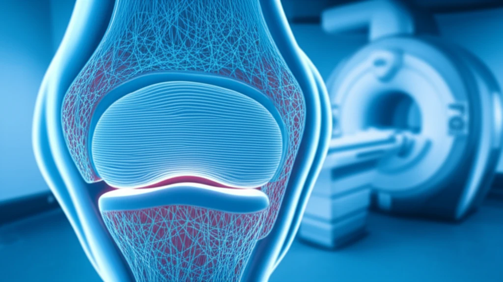
The temporomandibular joint (TMJ), where your jawbone connects to your skull, is a complex structure of bone, cartilage, and a shock-absorbing disc. This disc, made of tough fibrocartilage, is critical for smooth jaw movement, but its intricate internal structure has been difficult to study.

Now, groundbreaking research is shedding light on the TMJ disc's hidden architecture. By combining advanced 3-Tesla magnetic resonance imaging (MRI) with detailed microscopic analysis, scientists have created a high-resolution map of the collagen fiber network within the disc. This new understanding could revolutionize how we diagnose and treat TMJ disorders.

What's the Big Deal About Collagen in My Jaw?

Illustration of a human jaw joint with glowing collagen fibers showing aligned and random patterns. MRI scanner in the background.

Collagen, the most abundant protein in your body, acts like scaffolding, providing strength and structure to tissues. In the TMJ disc, collagen fibers are arranged in a specific network to withstand forces from chewing, talking, and other movements. This network isn't uniform; it varies across different regions of the disc.

Researchers used a powerful 3-Tesla MRI to scan the TMJs of cadaver heads, followed by a detailed microscopic analysis of the disc tissue. This combined approach allowed them to compare the MRI findings with the actual arrangement of collagen fibers. Here's what they discovered:

  • Central Zone: Collagen fibers are primarily aligned in a sagittal direction (front to back), providing strength along the main plane of movement.
  • Anterior and Posterior Zones: Fibers are arranged more randomly (isotropically), allowing for flexibility and shock absorption from various angles.
  • MRI Connection: MRI T2 values, which reflect water movement within the tissue, correlate with the degree of collagen fiber alignment. Higher T2 values were found in areas with more random fiber arrangements.
This is the first time scientists have systematically compared MRI data with the actual microscopic structure of the TMJ disc. The findings suggest that MRI can be a valuable tool for non-invasively assessing the health and structure of this critical joint.

What Does This Mean for My Jaw?

This research opens the door to more precise and personalized treatments for TMJ disorders. By using MRI to assess the collagen fiber network in your TMJ disc, doctors may be able to:

<ul> <li><b>Identify the specific structural problems</b> contributing to your pain and dysfunction.</li> <li><b>Develop targeted therapies</b> to address these problems, such as physical therapy to improve fiber alignment or regenerative medicine to repair damaged tissue.</li> <li><b>Monitor the effectiveness of treatments</b> over time using non-invasive MRI scans.</li> </ul>

While more research is needed, this study represents a significant step forward in our understanding of the TMJ. It highlights the importance of collagen fiber arrangement in joint health and suggests that MRI can be a powerful tool for diagnosing and treating TMJ disorders. If you're struggling with jaw pain, talk to your doctor about whether advanced imaging techniques could help you find relief.

About this Article -

This article was crafted using a human-AI hybrid and collaborative approach. AI assisted our team with initial drafting, research insights, identifying key questions, and image generation. Our human editors guided topic selection, defined the angle, structured the content, ensured factual accuracy and relevance, refined the tone, and conducted thorough editing to deliver helpful, high-quality information.See our About page for more information.

This article is based on research published under:

DOI-LINK: 10.11607/ofph.1879, Alternate LINK

Title: Regional Collagen Fiber Network In The Articular Disc Of The Human Temporomandibular Joint: Biochemical 3-Tesla Quantitative Magnetic Resonance Imaging Compared To Quantitative Histologic Analysis Of Fiber Arrangement

Subject: Anesthesiology and Pain Medicine

Journal: Journal of Oral & Facial Pain and Headache

Publisher: Quintessence Publishing

Authors: Jaryna Eder, Zbynek Tonar, Martina Schmid-Schwap, Margit Bristela, Astrid Skolka, Hannes Traxler, Eva Piehslinger, Monika Egerbacher, Siegfried Trattnig, Kirsti Witter

Published: 2018-07-01

Everything You Need To Know

1

What are temporomandibular joint disorders (TMD), and what makes them so difficult to treat?

Temporomandibular joint disorders (TMD) affect millions and are characterized by pain, limited movement, and a reduced quality of life. The causes are complex making effective treatment challenging. This new research using 3-Tesla MRI scans with microscopic analysis, aims to better understand the temporomandibular joint (TMJ) to make more effective treatments.

2

What role does collagen play in the jaw's temporomandibular joint (TMJ), and how is it structured differently in various parts of the joint?

Collagen acts as scaffolding in the body, providing strength and structure to tissues. In the TMJ disc, collagen fibers are arranged to withstand forces from chewing and talking. The arrangement of this network varies; collagen fibers in the central zone are aligned in a sagittal direction, providing strength along the main plane of movement. In the anterior and posterior zones, fibers are arranged more randomly, allowing for flexibility and shock absorption.

3

How did researchers combine 3-Tesla MRI and microscopic analysis to study the TMJ disc?

Researchers combined 3-Tesla magnetic resonance imaging (MRI) with microscopic analysis of the TMJ disc tissue in cadaver heads. They used the MRI to scan the TMJs and then conducted a detailed microscopic analysis of the disc tissue. This allowed them to compare MRI findings with the actual arrangement of collagen fibers.

4

What are MRI T2 values in the context of temporomandibular joint (TMJ) imaging, and how do they relate to collagen fiber arrangement?

MRI T2 values reflect water movement within the TMJ tissue and correlate with the degree of collagen fiber alignment. Higher T2 values were found in areas with more random fiber arrangements. This suggests that MRI can be a valuable tool for non-invasively assessing the health and structure of the TMJ.

5

What are the potential implications of this TMJ research for diagnosing and treating jaw disorders in the future?

The research findings suggest that MRI could be used to assess the collagen fiber network in the TMJ disc, which may allow doctors to diagnose problems earlier. This would allow for more precise and personalized treatments of TMJ disorders. This approach focuses on understanding the biomechanical properties of the joint, not just addressing the symptoms, which could lead to better long-term outcomes.

Newsletter Subscribe

Subscribe to get the latest articles and insights directly in your inbox.