
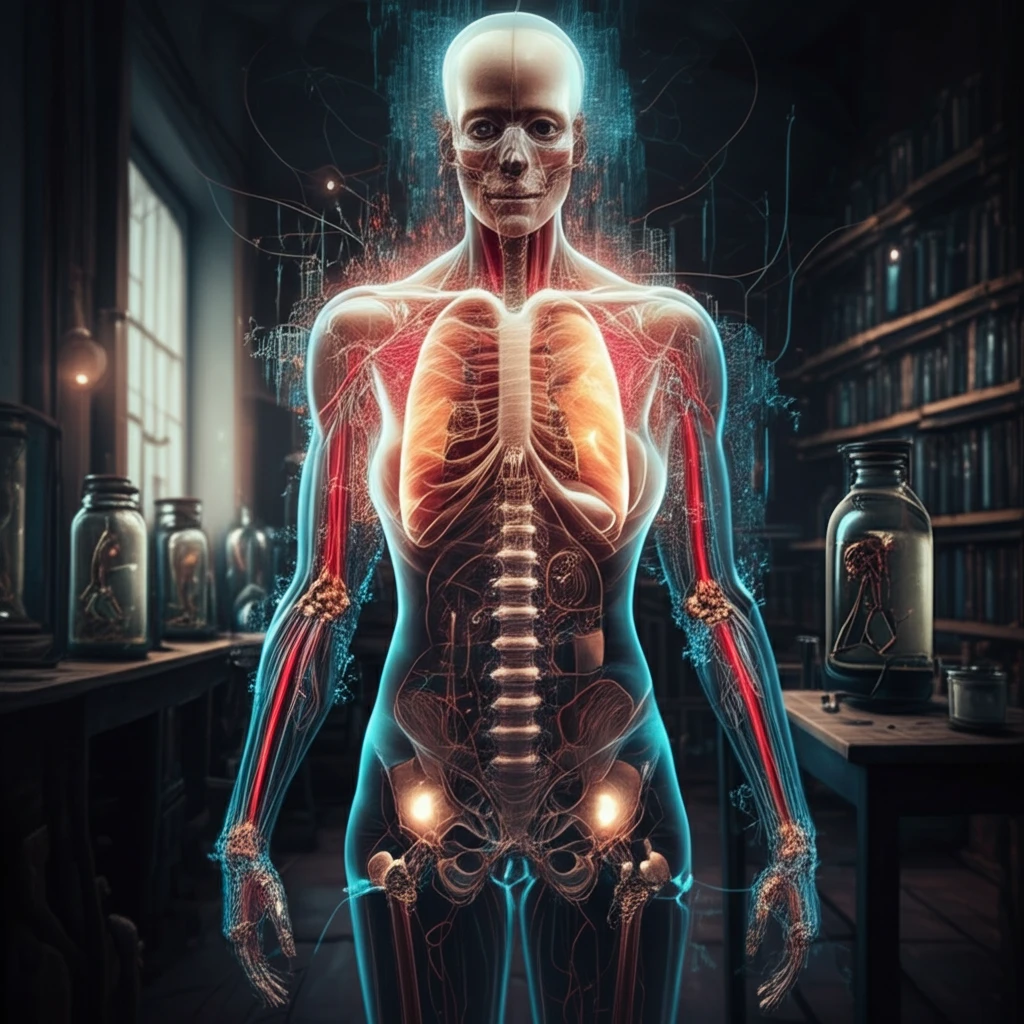
Unlocking the Past: How Digital Anatomy Preservation is Revolutionizing Medical Education and Heritage

"From ancient anatomical collections to cutting-edge 3D models, explore how technology is transforming the way we study the human body and preserve invaluable medical knowledge."
For centuries, the study of anatomy has been fundamental to medical education and practice. Anatomical collections, often painstakingly assembled and preserved, represent a tangible link to the past, offering invaluable insights into the intricacies of the human body. However, these collections are inherently fragile and vulnerable to damage or decay. Traditional methods of preservation, while effective to a degree, cannot fully prevent the gradual degradation of these irreplaceable resources.
Enter the digital revolution. In recent years, advancements in imaging technologies, 3D modeling, and virtual reality have opened up exciting new possibilities for preserving and studying anatomical specimens. Digital anatomy preservation offers a non-destructive means of capturing the intricate details of these historical artifacts, creating virtual replicas that can be studied, shared, and interacted with in ways never before imagined. This approach not only safeguards these precious collections for future generations but also enhances their accessibility and utility for medical education and research.
This article delves into the fascinating world of digital anatomy preservation, exploring its techniques, applications, and potential impact on medical education and our understanding of the human body. We will journey from the anatomical laboratories of Dakar to the cutting-edge imaging facilities of Montpellier, uncovering how technology is breathing new life into historical collections and revolutionizing the way we learn about anatomy.
Digital Platforms Transforming Anatomy Access
Digital anatomy platforms are expanding access to anatomical knowledge beyond traditional lab settings. The BioDigital Human platform offers interactive 3D visualizations of anatomy, disease, and treatment that can be embedded into any learning management system or digital solution. Meanwhile, projects like Anatomy Steward operate as focused digital museums dedicated to osteology and comparative anatomy, making preserved specimens accessible to broader audiences. These platforms represent a shift toward democratizing anatomical education through digital preservation and interactive visualization.
Traditional Preservation Methods and Their Drawbacks
Before modern chemical fixatives were introduced, the primary agents for anatomical preservation were alcoholic solutions of arsenic and alumina salts in varying concentrations. The later adoption of carbolic acid (phenol) and formaldehyde became the standard approach for embalming and specimen preservation, though these methods have well-documented limitations. Anatomical specimens remain subject to gradual tissue deterioration, and institutions face significant spatial and financial constraints in maintaining physical collections. These ongoing concerns about conservation and resource limitations have motivated the search for digital alternatives to traditional preservation.
From Ancient Drawings to Plastination
The history of anatomical illustration spans over 2,500 years, beginning with ancient Egyptian medical diagrams and progressing through medieval manuscripts to today's 3D digital models. A pivotal milestone in physical preservation came in 1977 when Gunther von Hagens developed plastination, a technique that replaces water and fat in body parts with plastics, yielding specimens that can be touched, do not smell, and resist decay. These milestones—from hand-drawn anatomical studies to polymer-preserved specimens—illustrate a long trajectory of innovation driven by the need to make anatomy tangible and enduring for learners.
The Rise of Digital Anatomy Preservation
Digital anatomy preservation encompasses a range of techniques aimed at capturing and replicating anatomical specimens in a digital format. These methods offer several key advantages over traditional preservation techniques: non-destructive imaging allows for detailed analysis without risking damage to the original specimen; digital models can be easily shared and accessed remotely, expanding access to valuable resources; and virtual reality and 3D printing technologies enable interactive exploration and physical replication of anatomical structures.
- Non-destructive: Digital techniques allow for study without damaging specimens.
- Accessibility: Digital models can be shared globally, democratizing education.
- Interactive: VR and 3D printing enable hands-on learning.
- Comprehensive: Capture both external and internal anatomical structures.
Advances in Adaptive Preservation and Interactive Learning
Recent research has introduced adaptive anatomical preservation optimal denoising (AAPOD), an innovative method designed to maximally reduce noise in daily radiation therapy MRI while preserving tissue boundaries. This technique demonstrates how computational approaches can maintain anatomical fidelity even under the constraints of repeated imaging. In parallel, platforms such as AnatomyLearning.com provide interactive 3D human anatomy visualization designed for students, health professionals, and educators, bridging the gap between research-level imaging and classroom instruction. Together, these developments highlight how digital tools are advancing both the precision of anatomical data and its accessibility for education.
Challenges in Chemical Preservation and Standardization
Efforts to improve anatomical tissue preservation through chemical means have encountered persistent challenges. Research into preservative blends—combining acid, buffer solutions, and cross-linking agents as mildew preventives, stabilizers, and fixatives respectively—has shown that characterizing and applying these mixtures to animal and human tissue remains complex. The field continues to grapple with establishing standardized preservation indices, as reflected in ongoing work on the Anatomical Preservation Index. These difficulties underscore that no single chemical method has fully solved the problem of long-term anatomical specimen integrity.
Comparing Preservation Approaches in Surgical Contexts
Comparative platforms such as Versus.com enable side-by-side evaluation of products and methods across over 100 categories using detailed specifications and data visualizations. In surgical contexts, maximum periprostatic anatomy-preserving robotic-assisted laparoscopic radical prostatectomy (MPAP-RALP) exemplifies how preservation-focused techniques are evaluated against conventional approaches. The emphasis on preserving anatomical structures during surgery mirrors the broader goal in anatomical science of maintaining tissue integrity, whether for patient outcomes or educational specimens. These comparisons highlight that preservation philosophy extends from the laboratory to the operating room.
The Future of Anatomy Education
Digital anatomy preservation is poised to revolutionize medical education, providing students with unprecedented access to anatomical resources and interactive learning experiences. Virtual dissection tables, powered by high-resolution digital models, allow students to explore the human body in a risk-free and engaging environment. These tables can be used to simulate surgical procedures, practice anatomical identification, and explore complex anatomical relationships. Furthermore, the ability to 3D print anatomical structures enables students to create physical models for hands-on study and practice. As technology continues to advance, digital anatomy preservation will play an increasingly important role in shaping the future of medical education, preparing future generations of healthcare professionals with a deeper understanding of the human body.
Expertise Gaps in Digital Preservation
Digital preservation demands specialized knowledge and skills that many institutions currently lack. Effective strategies require managing metadata, emulation, and migration of digital assets—tasks that go well beyond traditional archival practices. In anatomy education, institutions such as Swiss medical schools have reported improved outcomes by integrating cadaver-based gross anatomy more effectively into surgical curricula, suggesting that digital and physical approaches must complement each other. Bridging the expertise gap is essential for ensuring that digital anatomical resources are preserved and utilized to their full potential.
Digital Surrogates and Emerging Preservation Technologies
In November 2024, the National Endowment for the Humanities awarded a Research and Development grant to the University of Minnesota to create digital surrogates of often-fragile books with multi-layer flaps, such as historical flap anatomies, preserving their interactive nature for digital users. This project illustrates a frontier where physical artifacts are translated into interactive digital formats without losing their pedagogical function. As digital resources portals for the humanities and social sciences expand, anatomical preservation is poised to benefit from these cross-disciplinary advances in digitization and interactive modeling.
Preservation Across Disciplines and Deep Time
The history of anatomy spans millennia, from ancient Egyptian mummification practices to modern imaging technologies, each era bringing new methods of preserving and understanding the body. In paleontology, researchers have used X-ray imaging to create three-dimensional reconstructions of trilobite anatomy preserved by rapid burial in volcanic ash, revealing fine structural details through mineral-mediated fossilization. These cross-disciplinary parallels—from human tissue preservation to fossilized invertebrates—demonstrate that the challenge of preserving biological structure is universal, and that 3D imaging is emerging as a shared tool across fields.
Soft-Tissue Preservation Reshapes Scientific Understanding
A landmark study published in Science documented the preservation of soft-tissue blood vessels and cellular structures in a Tyrannosaurus rex specimen, challenging long-held assumptions about the limits of biological preservation over geological timescales. This discovery demonstrated that under specific conditions, delicate anatomical structures can endure for tens of millions of years, opening new avenues for studying ancient biology at the cellular level. The findings have profound implications not only for paleontology but also for our understanding of preservation science itself, suggesting that nature can achieve what laboratory methods still struggle to replicate.