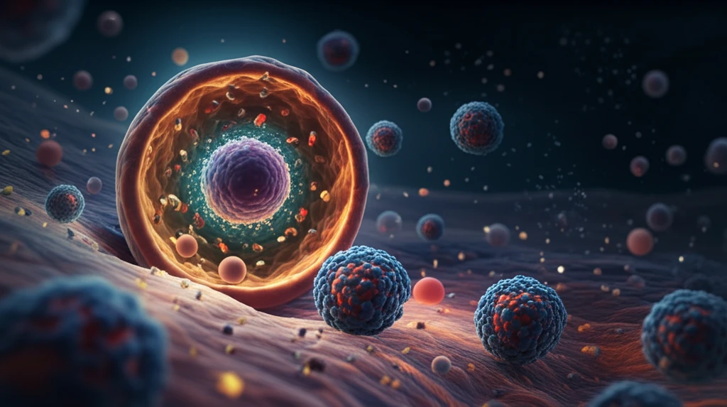
Surreal illustration of nanoparticles degrading inside a cell.

The Tiny Avengers: How Nanoparticles Are Revolutionizing Medicine from Within

"Unlocking the Secrets of Biodegradable Nanoparticles for Targeted Drug Delivery"


In the ever-evolving landscape of medical science, the development of smart drug delivery systems has captured the imagination of researchers worldwide. At the heart of this revolution lies biodegradable polymeric nanoparticles, microscopic vehicles capable of ferrying medications directly to diseased cells. This approach promises to minimize side effects and maximize therapeutic impact. Imagine tiny, biocompatible containers, loaded with life-saving drugs, navigating the intricate pathways within our bodies to precisely target tumors or repair damaged tissues.

Among the most promising materials for these nanoparticles is poly(L-lactic acid) (PLLA), a biodegradable polymer that has been extensively studied for its compatibility with biological systems. While the degradation of PLLA in various environments is well-documented, what happens when these nanoparticles enter individual cells remains a topic of intense scientific curiosity. How do they break down? What is the fate of the released drug? Answering these questions is crucial to unlocking the full potential of PLLA nanoparticles in medicine.

This article explores groundbreaking research into the intracellular degradation of PLLA nanoparticles, offering a glimpse into their fate within living cells. By tracking these particles and their components over time, scientists are gaining valuable insights that could pave the way for more effective and targeted therapies.

A Nanoparticle's Journey: Tracking PLLA Degradation Inside Cells

Surreal illustration of nanoparticles degrading inside a cell.

To observe the intracellular behavior of PLLA nanoparticles, scientists at the Max Planck Institute for Polymer Research designed a clever experiment. They created PLLA nanoparticles with an average diameter of approximately 120 nanometers and decorated them with magnetite nanocrystals. These nanocrystals acted as markers, allowing the researchers to track the nanoparticles' location and breakdown within mesenchymal stem cells (MSCs).

MSCs, a type of adult stem cell with regenerative properties, were chosen as a model cellular system. The researchers introduced the magnetite-studded PLLA nanoparticles into the MSCs and then meticulously monitored their fate using transmission electron microscopy (TEM) over a period of 14 days. TEM, a powerful imaging technique, provided detailed, high-resolution snapshots of the nanoparticles' journey inside the cells.

  • Magnetite as a Marker: The magnetite nanocrystals served as a visual cue, revealing when the PLLA nanoparticles began to degrade and release their contents.
  • Long-Term Monitoring: Observing the nanoparticles for two weeks allowed the researchers to capture both early and late stages of degradation.
  • High-Resolution Insights: TEM provided ultrastructural details, revealing how the cells processed the nanoparticles.
The TEM images revealed a fascinating story. As the PLLA nanoparticles resided within the MSCs, the magnetite nanocrystals began to detach from their surface, indicating that the PLLA was indeed undergoing degradation. Even after 14 days, remnants of the PLLA nanoparticles could still be found within the cells, suggesting that the degradation process was gradual and sustained. This observation highlights the potential of PLLA nanoparticles for long-term drug release.

The Future of Nanomedicine: Targeted Therapies and Beyond

This research underscores the importance of TEM studies in understanding the intracellular fate of nanoparticles. By combining TEM with other techniques like flow cytometry and confocal laser scanning microscopy (CLSM), scientists can gain a comprehensive picture of how cells interact with these tiny vehicles. These insights are crucial for designing more effective and targeted drug delivery systems. As our understanding of nanoparticle behavior within cells deepens, we can anticipate a future where nanomedicine plays an increasingly vital role in treating diseases and improving human health.

About this Article -

This article was crafted using a human-AI hybrid and collaborative approach. AI assisted our team with initial drafting, research insights, identifying key questions, and image generation. Our human editors guided topic selection, defined the angle, structured the content, ensured factual accuracy and relevance, refined the tone, and conducted thorough editing to deliver helpful, high-quality information.See our About page for more information.

This article is based on research published under:

DOI-LINK: 10.3762/bjnano.5.201, Alternate LINK

Title: Imaging The Intracellular Degradation Of Biodegradable Polymer Nanoparticles

Subject: Electrical and Electronic Engineering

Journal: Beilstein Journal of Nanotechnology

Publisher: Beilstein Institut

Authors: Anne-Kathrin Barthel, Martin Dass, Melanie Dröge, Jens-Michael Cramer, Daniela Baumann, Markus Urban, Katharina Landfester, Volker Mailänder, Ingo Lieberwirth

Published: 2014-10-29

Everything You Need To Know

1

What are biodegradable polymeric nanoparticles, and how are they revolutionizing drug delivery?

Biodegradable polymeric nanoparticles are microscopic vehicles designed to transport medications directly to diseased cells. This targeted approach aims to reduce side effects and enhance the therapeutic impact of drugs by delivering them precisely where they are needed in the body. The primary goal is to improve the effectiveness of treatments while minimizing harm to healthy tissues. Research into these nanoparticles is focused on understanding their behavior within the body, particularly how they interact with cells and release their drug payloads.

2

What methods do scientists employ to monitor the journey of nanoparticles within cells?

Scientists use techniques like transmission electron microscopy (TEM), flow cytometry, and confocal laser scanning microscopy (CLSM) to track nanoparticles within cells. In the specific study, transmission electron microscopy was crucial as it provided high-resolution images showing the location and degradation of poly(L-lactic acid) nanoparticles inside mesenchymal stem cells. Visual markers, like magnetite nanocrystals attached to the nanoparticles, help monitor their breakdown and the release of their contents.

3

Why is poly(L-lactic acid) (PLLA) considered a promising material for creating nanoparticles in drug delivery systems?

Poly(L-lactic acid) (PLLA) is a biodegradable polymer widely studied for its compatibility with biological systems and its ability to degrade over time. The study mentioned used PLLA to create nanoparticles for drug delivery, which makes it an excellent option for targeted drug delivery. The gradual breakdown of PLLA allows for sustained release of medication, potentially improving treatment outcomes. Understanding the intracellular degradation process of PLLA is key to optimizing its use in nanomedicine.

4

What did the research reveal about the degradation timeline of poly(L-lactic acid) nanoparticles within cells?

The research highlighted the gradual degradation of poly(L-lactic acid) nanoparticles inside mesenchymal stem cells (MSCs) over a 14-day period. Even after two weeks, remnants of the nanoparticles were still present, indicating a sustained release of the drug. This observation suggests that PLLA nanoparticles can provide long-term drug delivery, which is particularly beneficial for treatments requiring prolonged medication exposure. Continuous monitoring using transmission electron microscopy allowed researchers to track this process in detail.

5

How can the insights gained from tracking nanoparticles impact the future of nanomedicine and targeted therapies?

The knowledge gained from tracking the intracellular fate of nanoparticles, particularly poly(L-lactic acid) nanoparticles, can be applied to design more effective and targeted drug delivery systems. For example, understanding the degradation rate and pathway of PLLA within cells can help optimize the release of therapeutic agents. This knowledge contributes to the advancement of nanomedicine, potentially leading to improved treatments for various diseases and enhanced human health through more precise and efficient therapies.

Newsletter Subscribe

Subscribe to get the latest articles and insights directly in your inbox.