
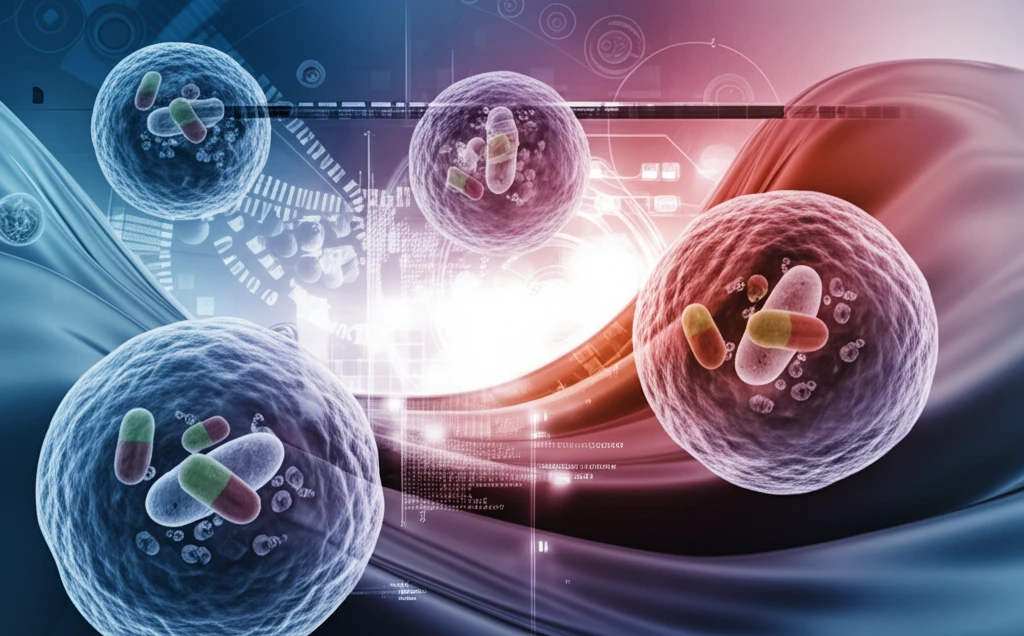
Nanoparticle Imaging: Unveiling the Secrets of Liposomes with Advanced Microscopy Techniques

"A Deep Dive into Transmission Electron Microscopy (TEM) and Its Revolutionary Role in Visualizing Nanoscale Drug Delivery Systems"
In the rapidly evolving field of nanotechnology, liposomes have emerged as promising vehicles for targeted drug delivery and diagnostic applications. These tiny, spherical vesicles, composed of lipid bilayers, can encapsulate and transport therapeutic agents directly to diseased cells, minimizing side effects and maximizing treatment efficacy. However, to fully harness the potential of liposomes, scientists must be able to precisely characterize their size, shape, and internal architecture.
Enter transmission electron microscopy (TEM), a powerful technique that allows researchers to visualize nanoparticles at the atomic level. TEM uses a beam of electrons to illuminate a sample, creating high-resolution images that reveal intricate details of liposome structure. This capability is crucial for optimizing liposome design, understanding drug encapsulation mechanisms, and ensuring the quality and consistency of nanoparticle formulations.
While imaging metal particles using electron microscopy is straightforward due to their high density and stability, visualizing soft material nanoparticles like liposomes requires specialized techniques to preserve their delicate structure. This article explores the cutting-edge methods employed in TEM to image liposomes, including cryo-electron microscopy (cryo-EM) and negative staining TEM, and discusses their respective advantages and limitations.
Explosive Growth in Nanomedicine
Multifunctional liposomes for imaging-guided therapy have enjoyed explosive development in nanomedicine, driven by the exciting ability to combine diagnostic and/or therapeutic agents into a single agent. The incorporation of nanoparticles into liposomes has gained particular interest recently due to the potential for enhancement. Most recent advances in the field also include the study of liposomes as synthetic cell mimics.
Choosing the Right Imaging Technique
A plethora of techniques for the imaging of liposomes and other bilayer vesicles are available, yet sample preparation and the technique chosen should be carefully considered in conjunction with the information required. Several imaging techniques are currently available for evaluating the morphology of liposomes and other nanoparticles, with each having its own advantages and disadvantages that should be considered when interpreting results.
From Electron Beams to Vesicle Imaging
Electron microscopy is a technique in which a beam of electrons is used as a source of illumination, and transmission electron microscopy transmits that beam through a specimen to form an image. These foundational principles underpin the modern imaging approaches used to characterize liposomes and other bilayer vesicles at high resolution.
Advanced TEM Techniques for Liposome Imaging
Transmission electron microscopy (TEM) is invaluable for characterizing the size and shape of nanoparticles, offering direct visualization of individual particles and their internal architecture. When imaging soft materials like liposomes, preserving their structure is essential. Cryo-electron microscopy (cryo-EM) is the best method for visualizing liposomes close to their native structure. In cryo-EM, thin films of suspensions are rapidly frozen to create vitrified ice films, which are then imaged directly in the electron microscope at liquid nitrogen temperatures.
- Cryo-Electron Microscopy (Cryo-EM): Best for preserving native structure, involves rapid freezing and imaging at cryogenic temperatures.
- Negative Staining TEM: Faster and simpler, uses heavy metal salts for contrast but may introduce artifacts.
- Freeze Fracture: Useful for larger liposomes, involves fracturing the sample and metal shadowing.
- Atomic Force Microscopy (AFM): Provides complementary information on liposome structure and mechanical properties.
Advances in Molecular and Multifunctional Imaging
The use of liposomes in molecular imaging makes it possible to image the alterations of different disease states, offering an imaging procedure that is acceptable for both physicians and especially child patients with faster metabolic activity. Acoustic liposomes, which are made of perfluoropropane gas, can be used as contrast agents in ultrasound imaging, while the encapsulation of quantum dots and fluorescent dyes into liposomes has led to further labeling strategies.
Limitations and Trade-Offs
Although a number of imaging techniques exist for evaluating liposome morphology, each has its own advantages and disadvantages that should be considered when interpreting results. Sample preparation and the choice of technique must be weighed against the specific information required, underscoring that no single method is universally ideal for all liposome characterization questions.
Negative Staining Versus Cryo-Electron Microscopy
The best method to visualize liposomes close to their native structure is cryo-electron microscopy, where thin films of suspensions are plunge frozen to create vitrified ice films that can be imaged directly in the electron microscope. By contrast, negative staining transmission electron microscopy offers an alternative route, though the two approaches differ substantially in sample preparation and in how faithfully they preserve vesicle morphology.
The Future of Liposome Imaging
As nanotechnology continues to advance, the demand for high-resolution imaging techniques will only increase. Cryo-EM, with its ability to visualize liposomes in their native state, is poised to become the gold standard for liposome characterization. Ongoing developments in TEM technology, such as improved detectors and automated data acquisition, are further enhancing the capabilities of liposome imaging. These advancements will not only deepen our understanding of liposome structure and function but also accelerate the development of more effective and targeted drug delivery systems for a wide range of diseases.
Matching Technique to Question
The choice among the many available imaging techniques for liposomes should be made carefully in conjunction with the information required, since each method carries distinct advantages and limitations. Cryo-electron microscopy is favored for viewing vesicles close to their native structure, positioning it as a reference approach for structural studies of liposomes and other bilayer vesicles.
Imaging-Guided Therapy and Synthetic Cells
Multifunctional liposomes that combine diagnostic and/or therapeutic agents into a single agent point toward imaging-guided therapy as a major frontier in nanomedicine. Recent advances exploring liposomes as synthetic cell mimics, together with the incorporation of nanoparticles into liposomes for potential enhancement, suggest active new directions for research.
Interpreting Morphology Across Techniques
Interpreting liposome morphology requires recognizing that the available imaging techniques each have their own advantages and disadvantages that influence results. Standardization across methods and careful control of sample preparation remain systemic challenges for reproducible characterization of nanoparticles.
Friendlier Imaging for Patients
Liposome-based molecular imaging offers a procedure that is more acceptable for both physicians and especially child patients with faster metabolic activity. By enabling the imaging of alterations in different diseases, these approaches support earlier and less invasive diagnosis.