Regenerating spinal cord with glowing nerve fibers

Spinal Cord Breakthrough: Can FGF1 and Nerve Grafts Restore Movement?

"New research explores how a biodegradable device delivering FGF1 and nerve grafts could revolutionize spinal cord injury treatment and motor function recovery."


Spinal cord injury (SCI) presents a formidable challenge in the field of neuroscience. While numerous strategies have emerged in recent years, aimed at repairing the central nervous system (CNS), the quest for effective treatments continues. The focus often lies in optimizing post-injury conditions, mitigating secondary damage, and fostering regeneration across the injury site to create a more favorable environment.

Recent breakthroughs have sparked renewed hope, particularly for individuals with chronic SCI. Epidural stimulation of the caudal spinal cord, combined with locomotor training, has shown promise in enabling voluntary movements of the lower extremities. This approach is believed to activate previously dormant axons that traverse the lesion. There is a theory in the scientific community, that even in cases of complete SCI, a regeneration strategy combined with locomotor training and epidural stimulation could potentially restore movement in the lower extremities.

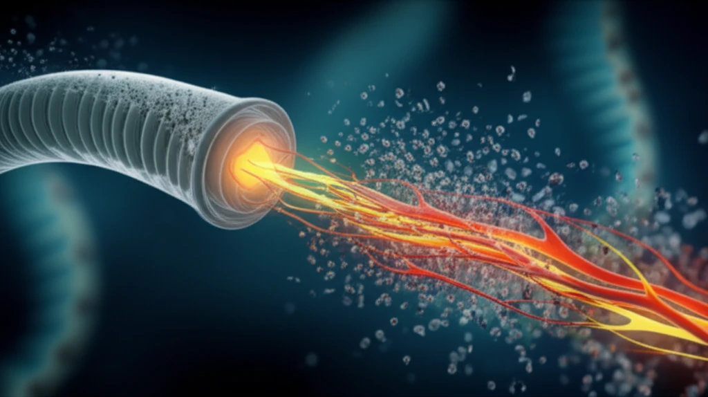
Peripheral nerve grafts (PNGs) present a potential avenue for regaining function in injured spinal cords. Some approaches combine PNGs with acidic fibroblast growth factor (FGF1), employing a strategy of rerouting regenerating fibers from white matter into adjacent gray matter. Studies suggest that longitudinal tracts tend to favor growth into gray matter, irrespective of guidance cues. Precise placement of PNGs is crucial for achieving optimal axonal growth.

PNGs and FGF1: A Cutting-Edge Strategy for Spinal Cord Repair?

Regenerating spinal cord with glowing nerve fibers

Researchers explored the use of a biodegradable device containing PNGs, with and without FGF1, to repair spinal cord injuries. This new device is made of calcium sulphate, designed to biodegrade, and slowly release FGF1. The study aimed to determine if the new device containing autologous PNGs could trigger a motor cortex-derived electrophysiological response in the hind limb (MEP), and whether the addition of FGF1 would enhance this response, with motor cortex derived hind limb response paralleled by motor cortex neuron regeneration across the injury zone.

Adult female Sprague Dawley rats were subjected to a T11 spinal cord resection, followed by repair using twelve white-to-grey matter oriented PNGs pre-positioned in a biodegradable device with or without FGF1. The rats were then evaluated using several methods:

  • BBB score (Basso, Beattie, and Bresnahan locomotor rating scale) to assess motor function.
  • Electrophysiology to measure motor evoked potentials (MEPs).
  • Immunohistochemistry, including anterograde BDA tracing, to analyze nerve fiber regeneration.
The results indicated that motor evoked potentials (MEPs) in the lower limb reappeared at 20 weeks post-grafting. The reappearance of MEPs was enhanced in the group treated with adjuvant FGF1. This improvement was paralleled by NF-positive fibers and anterogradely traced corticospinal fibers distal to the injury. The BBB scores also showed improvements in the repaired animals. This research supports the idea that combining PNGs and FGF1 can be a regeneration strategy to reinnervate the caudal spinal cord. The new device induced robust MEPs augmented by FGF1, and may be considered for translational research.

Looking Ahead: The Future of Spinal Cord Injury Treatment

These findings offer valuable insights into potential therapeutic strategies for spinal cord injuries. The combination of PNGs and FGF1, facilitated by a biodegradable device, represents a promising approach to promote nerve regeneration and restore motor function. While further research is needed, this study provides a strong foundation for future translational studies and potential clinical applications.

About this Article -

This article was crafted using a human-AI hybrid and collaborative approach. AI assisted our team with initial drafting, research insights, identifying key questions, and image generation. Our human editors guided topic selection, defined the angle, structured the content, ensured factual accuracy and relevance, refined the tone, and conducted thorough editing to deliver helpful, high-quality information.See our About page for more information.

Everything You Need To Know

1

What is the role of FGF1 in spinal cord injury repair, and how does it work with Peripheral Nerve Grafts (PNGs)?

FGF1, or acidic fibroblast growth factor, is a key element in promoting nerve regeneration after spinal cord injury. The research indicates that the addition of FGF1 to the biodegradable device containing PNGs enhanced the reappearance of Motor Evoked Potentials (MEPs). FGF1 is believed to support the growth and guidance of regenerating nerve fibers. This combined approach, using PNGs and FGF1, aims to reinnervate the caudal spinal cord, leading to improved motor function. The biodegradable device serves as a delivery system, ensuring the localized release of FGF1 at the injury site, which maximizes its regenerative effects.

2

How does the use of a biodegradable device contribute to the treatment of spinal cord injuries?

The biodegradable device plays a crucial role in delivering the therapeutic agents, specifically Peripheral Nerve Grafts (PNGs) and FGF1, directly to the site of the spinal cord injury. The device is made of calcium sulphate and designed to slowly release FGF1. This controlled release ensures a sustained presence of FGF1, supporting nerve regeneration over time. The device also facilitates the precise placement of PNGs, guiding the regenerating nerve fibers across the injury site. Its biodegradable nature means that it gradually dissolves, leaving behind the regenerated tissue without requiring a secondary removal procedure. This innovative approach aims to promote a favorable environment for nerve repair and motor function recovery.

3

What are Peripheral Nerve Grafts (PNGs), and why are they used in spinal cord injury repair?

Peripheral Nerve Grafts (PNGs) are essentially pieces of nerve tissue taken from other parts of the body and transplanted to the site of the spinal cord injury. They serve as a bridge, providing a pathway for nerve fibers to regrow across the damaged area. PNGs are used because they can reroute regenerating fibers from white matter into adjacent gray matter, which is crucial for restoring motor function. The use of PNGs, in conjunction with FGF1, creates a synergistic effect, maximizing the potential for nerve regeneration and functional recovery. The study mentioned the specific white-to-grey matter orientation of the PNGs.

4

What is the significance of Motor Evoked Potentials (MEPs) in assessing the success of spinal cord injury treatment?

Motor Evoked Potentials (MEPs) are electrophysiological responses that indicate the activation of motor pathways. Their reappearance after spinal cord injury repair signifies that the nerve signals are once again able to travel from the motor cortex to the hind limbs. The study measured motor evoked potentials (MEPs) in the lower limb. The enhanced MEPs in the group treated with FGF1 is a very important finding, indicating improved nerve fiber regeneration. The presence and improvement of MEPs serve as a direct measure of the effectiveness of the treatment in restoring the connection between the brain and the muscles, as well as a crucial step in restoring motor function and voluntary movements.

5

Beyond the specific methods discussed, what are the broader implications of this research for the future of spinal cord injury treatment?

The research, specifically the combination of Peripheral Nerve Grafts (PNGs), acidic fibroblast growth factor (FGF1), and a biodegradable device, holds significant implications for the future of spinal cord injury treatment. It represents a shift towards regenerative medicine, aiming not just to stabilize the injury but to actively repair the damaged spinal cord. These findings suggest that the restoration of movement and the potential to restore the spinal cord is within reach. It paves the way for translational studies and future clinical applications. The use of a biodegradable device for drug delivery and cell therapy is an innovative approach. Further research could explore refinements to the methods, patient specific solutions, and optimization of treatment parameters, which could greatly improve outcomes for individuals with spinal cord injuries.

Newsletter Subscribe

Subscribe to get the latest articles and insights directly in your inbox.