MRI scan showing a clear airway of a sleeping person

Snoring Isn't Always a Joke: How Imaging Can Help Diagnose Sleep Apnea

"Uncover the hidden causes of obstructive sleep apnea with advanced imaging techniques."


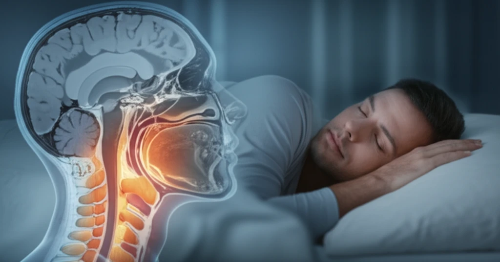
Obstructive sleep apnea (OSA) is more than just a nighttime annoyance; it's a condition characterized by repeated upper airway obstructions during sleep. These obstructions, occurring at the level of the pharynx, can lead to serious health issues if left unaddressed.

While traditional methods like cephalometric analysis (using X-rays) play a role in assessing craniofacial deformities related to OSA, modern imaging techniques like computed tomography (CT) and magnetic resonance imaging (MRI) are proving invaluable. These advanced tools allow doctors to visualize the anatomy of the upper airway in detail, helping them identify the specific causes of obstruction.

This article explores how CT and MRI scans are used to diagnose OSA, highlighting the key anatomical features that radiologists and doctors evaluate to create effective treatment plans.

Why Imaging Matters in Diagnosing OSA

MRI scan showing a clear airway of a sleeping person

OSA is often caused by a combination of factors, making it crucial to have a comprehensive understanding of the patient's anatomy. Both MRI and CT scans provide detailed images of the upper airway, allowing doctors to:

These imaging techniques offer several advantages:

  • Pinpoint the obstruction site: Identify exactly where the airway is collapsing or narrowing.
  • Assess soft tissue: Evaluate the size and shape of the tongue, soft palate, and other tissues that contribute to obstruction.
  • Evaluate bone structures: Examine the position of the jaw, hyoid bone, and other bony structures that influence airway patency.
  • Guide treatment decisions: Inform surgical planning and other interventions.
MRI is favored due to its ability to provide excellent anatomical resolution without using ionizing radiation. However, CT scans offer a quick and effective alternative, especially when bone structures are of primary interest.

What to Expect During Imaging

If your doctor suspects you have OSA, they may recommend an MRI or CT scan of your upper airway. Here's a general idea of what to expect:

<b>MRI:</b> You'll lie down on a table that slides into a large, tube-shaped machine. The MRI uses strong magnetic fields and radio waves to create detailed images. The scan can take anywhere from 30 to 60 minutes, and it's important to stay still during the process.

<b>CT Scan:</b> You'll lie on a table that slides into a donut-shaped machine. The CT scanner uses X-rays to create cross-sectional images of your airway. The scan is typically very quick, often taking just a few minutes.

About this Article -

This article was crafted using a human-AI hybrid and collaborative approach. AI assisted our team with initial drafting, research insights, identifying key questions, and image generation. Our human editors guided topic selection, defined the angle, structured the content, ensured factual accuracy and relevance, refined the tone, and conducted thorough editing to deliver helpful, high-quality information.See our About page for more information.

Everything You Need To Know

1

What exactly is Obstructive Sleep Apnea (OSA), and why is it important?

Obstructive Sleep Apnea (OSA) is a condition where the upper airway repeatedly becomes blocked during sleep, leading to pauses in breathing. These obstructions typically happen at the level of the pharynx. If left untreated, OSA can contribute to serious health problems. It's significant because it affects breathing during sleep, potentially leading to low oxygen levels and other health complications.

2

How do MRI and CT scans help in diagnosing OSA?

MRI and CT scans are advanced imaging techniques used to visualize the anatomy of the upper airway. They help doctors pinpoint the exact location of airway obstructions, assess soft tissues like the tongue and soft palate, and evaluate bony structures such as the jaw and hyoid bone. These scans offer detailed images that guide treatment decisions and surgical planning, providing a comprehensive understanding of the patient's anatomy related to OSA.

3

What are the main differences between MRI and CT scans for OSA, and which one is better?

MRI is often preferred because it provides excellent anatomical resolution without using ionizing radiation. This means there's no exposure to radiation during the scan. CT scans are a quick and effective alternative, particularly when the focus is on bone structures. The choice between MRI and CT depends on specific diagnostic needs and the structures doctors want to examine closely.

4

What can I expect during an MRI or CT scan if my doctor suspects I have OSA?

During an MRI or CT scan for OSA, you can expect to lie still while the scanner takes detailed images of your upper airway. These scans allow doctors to identify the exact location of the obstruction, evaluate soft tissues like the tongue and soft palate, and assess bone structures. The results inform treatment plans and guide interventions to address the underlying causes of OSA.

5

How do modern imaging techniques like CT and MRI compare to older methods like cephalometric analysis for diagnosing OSA?

Cephalometric analysis, using X-rays, is a traditional method used to assess craniofacial deformities related to OSA. However, modern imaging techniques like CT and MRI offer more detailed insights into the upper airway's anatomy. CT and MRI provide higher resolution images, enabling doctors to pinpoint obstruction sites, assess soft tissues, and evaluate bone structures more effectively. This leads to more accurate diagnoses and tailored treatment strategies compared to the traditional method.

Newsletter Subscribe

Subscribe to get the latest articles and insights directly in your inbox.