
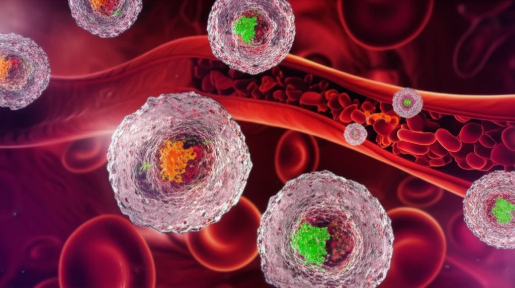
Sepsis-Induced Coagulopathy: How Extracellular Vesicles Turn Messengers into Mayhem

"Unlocking the role of extracellular vesicles (EVs) in the progression of sepsis-induced coagulopathy and its potential impact on therapeutic interventions."
Sepsis, a life-threatening condition arising from the body's overwhelming response to an infection, often leads to a dangerous complication known as sepsis-induced coagulopathy. This condition disrupts the normal blood clotting process, causing a cascade of events that can damage organs and increase the risk of death. For years, researchers have been working tirelessly to unravel the complexities of this process, seeking new ways to diagnose, prevent, and treat it effectively.
In recent years, a spotlight has been cast on tiny cellular structures called extracellular vesicles (EVs). These vesicles, once dismissed as mere cellular debris, are now recognized as key players in intercellular communication, acting as messengers that carry proteins, lipids, and genetic material between cells. The surge of research into EVs has captivated clinicians and researchers alike, offering new insights into the mechanisms driving coagulation disorders in sepsis.
This article aims to provide an updated overview of the latest knowledge on EVs and their involvement in sepsis-induced coagulopathy. We'll explore the structure and function of these vesicles, their pro-inflammatory and procoagulant properties, and their impact on the development of coagulation disorders and organ dysfunction. Furthermore, we'll discuss current challenges and future directions in this rapidly evolving field.
EVs and Sepsis Burden
Sepsis-induced disseminated intravascular coagulopathy remains a challenging clinical condition associated with significant morbidity and mortality. Tissue factor is the primary physiological initiator of the extrinsic coagulation pathway, and in Gram-negative sepsis, lipopolysaccharide activates monocytes, promoting tissue factor expression and release of tissue factor-bearing extracellular vesicles into the circulation. Enhanced caspase-1 activity and declining miR-126 levels in circulating extracellular vesicles are associated with sepsis-related organ failure and mortality. These vesicular contents may serve as prognostic biomarkers or future therapeutic targets.
Scoring Sepsis Coagulopathy
Sepsis-induced coagulopathy commonly begins with hypercoagulability, while a shift toward hypocoagulability generally occurs only in late sepsis after disseminated intravascular coagulation develops. Current diagnostic work applies the sepsis-induced coagulopathy score alongside the revised 2025 ISTH DIC score, with SIC intended to detect earlier-phase coagulopathy. Studies also measure extracellular vesicle tissue factor activity over time in plasma from patients with sepsis, including patients with or without DIC or SIC. Together, these approaches provide a more nuanced classification of coagulation status, but the need to distinguish early SIC from later DIC remains a central limitation.
An Unstandardized Foundation
By 2018, the basic concept that extracellular vesicles have procoagulant properties and may contribute to sepsis-induced coagulopathy had begun to emerge. However, the actual state of knowledge remained far from satisfactory. A major reason was the lack of standardized assay procedures, which limited confident comparison and interpretation of findings across studies.
Extracellular Vesicles: Structure and Function
Extracellular vesicles (EVs) are a diverse group of membrane-enclosed vesicles secreted by cells. These vesicles are categorized based on their size and biogenesis, with key types including apoptotic bodies, exosomes, and microvesicles. Apoptotic bodies, the largest EVs (0.5-5 μm), are formed during cellular disassembly, while exosomes (0.03-0.15 μm) originate from endocytosis and exocytosis. Microvesicles (0.1-5 μm) bud directly from the plasma membrane.
- Apoptotic Bodies: Large vesicles released during cell death.
- Exosomes: Small vesicles involved in cell-to-cell communication.
- Microvesicles: Shed from the plasma membrane.
EVs as Distributed Coagulant Signals
Recent reviews describe extracellular vesicles as having procoagulant properties during sepsis and as capable of disseminating prothrombotic activity by transferring those properties to distant target cells. Sepsis-associated hemostatic disorders can lead to extensive intravascular thrombosis, disseminated intravascular coagulation, multi-organ dysfunction syndrome, and increased mortality. Sepsis-induced coagulopathy is also characterized as arising from an uncontrolled acquired immunoinflammatory response and stimulation of coagulation mechanisms.
The Balance Is Context-Dependent
The role of endothelial cell-derived extracellular vesicles in the progression of sepsis-induced coagulopathy remains uncertain. Their apparent effects depend on the balance between procoagulant and anticoagulant activity, which can change according to the surrounding conditions. This context dependence limits any simple conclusion that endothelial extracellular vesicles are uniformly harmful or uniformly protective.
The Future of EVs in Sepsis Research
Recent studies have successfully illuminated the significant roles of EVs in the progression of coagulation disorders in sepsis. However, further harmonization in terminology, methodology, and evaluation methods is required for future studies. Standardized assays are crucial for accurate and comparable results. As we continue to unravel the complexities of EV function, we may discover new therapeutic targets to combat sepsis-induced coagulopathy and improve patient outcomes.