
VWF Unveiled: Comparing Recombinant vs. Plasma-Derived Forms for Enhanced Blood Clotting Therapies

"Atomic Force Microscopy reveals structural secrets, paving the way for improved von Willebrand factor treatments."
Von Willebrand factor (VWF) is a crucial protein in our blood, acting like a molecular glue that helps initiate blood clotting. Think of it as the first responder at the scene of an injury, attracting platelets to form a plug and stop the bleeding. Inherited deficiencies or defects in VWF lead to von Willebrand disease (VWD), a common bleeding disorder with symptoms ranging from mild to severe.
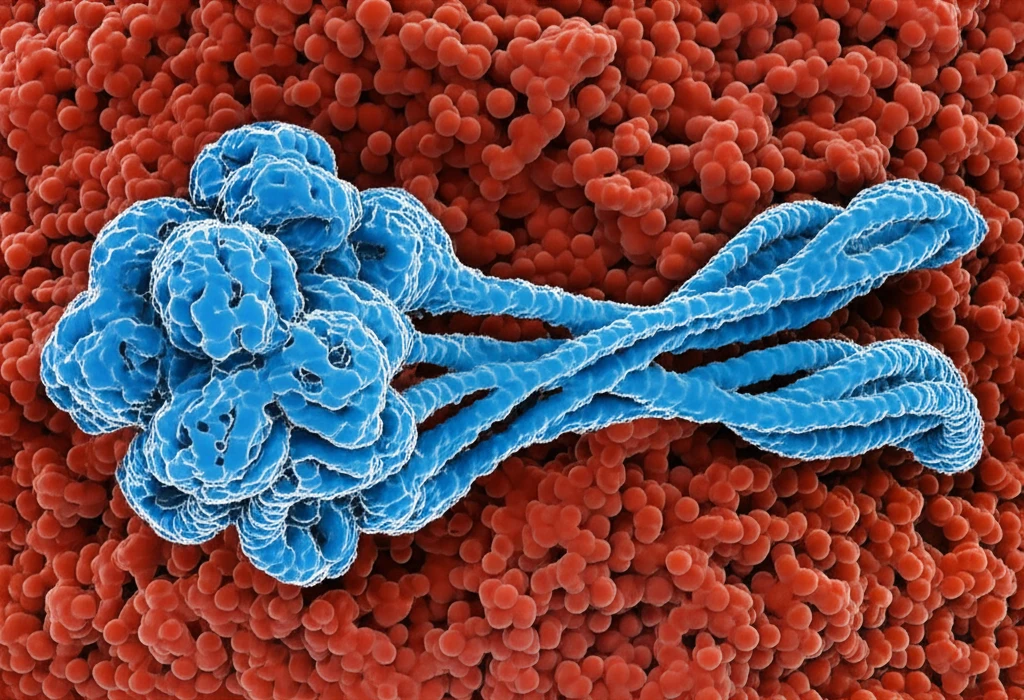
VWF comes in different forms, primarily sourced from blood plasma (pdVWF) or manufactured through recombinant technology (rVWF). Both aim to restore proper clotting function in VWD patients, but they aren't identical. A key difference lies in their structure, particularly the presence of ultra-large multimers, which are the most active forms of VWF. Understanding these structural nuances is vital for optimizing treatment strategies.
This article delves into the microscopic world of VWF, comparing the structural characteristics of pdVWF and rVWF using atomic force microscopy (AFM). We'll explore how these forms differ in size, shape, and behavior under stress, shedding light on potential implications for their effectiveness in treating bleeding disorders.
Clinical Importance of VWF
Von Willebrand factor is central to normal blood clotting, so abnormalities can contribute to clinically significant bleeding. The impact may vary substantially with the underlying defect and an individual's circumstances. Comparing recombinant and plasma-derived therapies therefore requires attention to both clotting support and patient-specific needs.
Diagnosis and Its Challenges
Diagnosis of von Willebrand disease can be difficult because the disorder is heterogeneous, laboratory assays have limitations, and physiologic processes can affect VWF measurements. CDC guidance notes that clotting-factor testing may show a longer clotting time in some people with VWD. Laboratory evaluation may also consider an increased VWF propeptide-to-antigen ratio when accelerated VWF clearance is suspected, while a streamlined approach could help optimize management in settings with limited resources and expertise.
A Century of Discovery
VWD was first described by Erik von Willebrand in 1926, making 2025 a centenary year for the disease. The factor associated with the prolonged bleeding time was subsequently named von Willebrand factor in his honor. Variant forms of VWF were recognized in the 1970s and are now understood as resulting from synthesis of an abnormal protein.
Peeking Under the Microscope: AFM Reveals VWF Structure
Atomic force microscopy (AFM) is a powerful tool that allows scientists to visualize molecules at the nanoscale. Imagine using a tiny finger to 'feel' the surface of a molecule, creating an image based on the texture and shape. In this study, researchers used AFM to compare the structures of pdVWF and rVWF, focusing on their lengths, the diameter of their globular domains (core units), and their behavior when exposed to shear stress (the force of blood flow).
- Length Distribution: Most VWF molecules, regardless of their origin (pdVWF or rVWF), fell within the 100-300 nm length range.
- Ultra-Large Multimers: Recombinant VWF (rVWF) had a slightly higher proportion of very long molecules (>300 nm) compared to pdVWF, suggesting a greater presence of ultra-large multimers.
- Globular Domain Size: The diameters of the globular core structures were similar for both types of VWF, ranging from 12 to 30 nm.
- Response to Shear Stress: Both pdVWF and rVWF undergo a dramatic conformational change when exposed to shear stress, extending their structure. This extension is crucial for VWF to function properly in initiating blood clotting.
Mechanisms Under Investigation
Recent work has identified a specific mechanism by which VWF binds platelets and initiates clotting under relevant circulatory conditions. Research using endothelialized fluidic devices found that aberrant flow patterns associated with venous valve pockets can produce VWF-platelet tangles resistant to ADAMTS13 removal. Reviews emphasize that VWF interacts with endothelial matrices, platelets, and factor VIII, linking primary hemostasis with coagulation.
Balancing Bleeding and Clotting
VWD is the most common bleeding disorder and can cause nosebleeds, easy bruising, heavy menstrual bleeding, and prolonged bleeding after injury, surgery, dental work, or childbirth. The condition may reflect insufficient VWF or defective VWF, making its pathophysiology important when interpreting diagnostic tests. At the same time, persistently high VWF or factor VIII levels can shift concern toward venous clotting risks such as deep vein thrombosis or pulmonary embolism.
Product Choice and Risk
Dosing recommendations for factor VIII concentrates containing VWF and recombinant VWF concentrates are individualized according to factors including body weight, VWD type, hemorrhage severity, FVIII and VWF levels, and the presence of inhibitors. VWF is also a multifaceted hemostatic protein whose interactions with endothelial matrices, platelets, and factor VIII affect both primary hemostasis and coagulation. Because increased VWF and FVIII levels are associated with increased venous thrombosis risk, comparison of products should consider both bleeding control and potential thrombotic concerns.
Implications for VWF Therapies
This microscopic comparison of pdVWF and rVWF provides valuable insights for the development and optimization of VWF therapies. The structural similarities between the two forms are reassuring, suggesting that rVWF can effectively mimic the function of its plasma-derived counterpart.
A Tailored Therapeutic Balance
The available material supports viewing VWF therapy as a balance between restoring effective hemostasis and managing individual risks. Product selection is unlikely to be meaningful without considering the patient's VWD characteristics, laboratory findings, and treatment context. Clinical decisions should therefore remain individualized and guided by specialist assessment.
Toward More Precise Care
Future progress may depend on improving diagnostic precision and matching VWF replacement strategies to distinct disease mechanisms. Research into VWF-platelet interactions and flow-sensitive clot formation could clarify how treatment affects hemostasis in different settings. These possibilities remain areas for continued investigation rather than established clinical conclusions.
VWF Beyond Hemostasis
Blood flow influences how VWF interacts with blood cells and shapes the characteristics of forming clots. VWF also functions as an inflammatory anchor for platelets at the blood-endothelium junction, linking endothelial breakdown and hematologic dysregulation to broader disease processes. In addition, studies report roles for VWF in venous thromboembolic disease and blood-vessel formation, while VWD itself includes both quantitative and qualitative defects in VWF.
Evidence From Clinical Practice
Real-world studies are examining how VWF therapies perform outside controlled trial settings. One multicenter, observational, retrospective study evaluated the efficacy and safety of plasma-derived VWF/FVIII-C products Fanhdi and Alphanate from Grifols at 10 Italian centers. Other reports describe treatment characteristics and outcomes among European patients receiving prophylaxis with recombinant or plasma-derived VWF, as well as US patients aged 12 years or older receiving routine or intermittent prophylactic recombinant VWF for menorrhagia.
The finding that rVWF may contain a higher proportion of ultra-large multimers is particularly interesting. These larger multimers are known to be more active in promoting blood clotting, potentially leading to improved therapeutic efficacy. However, further research is needed to fully understand the clinical implications of this difference.
Ultimately, a deeper understanding of VWF structure and function will pave the way for more targeted and effective treatments for von Willebrand disease, improving the lives of individuals affected by this common bleeding disorder.