
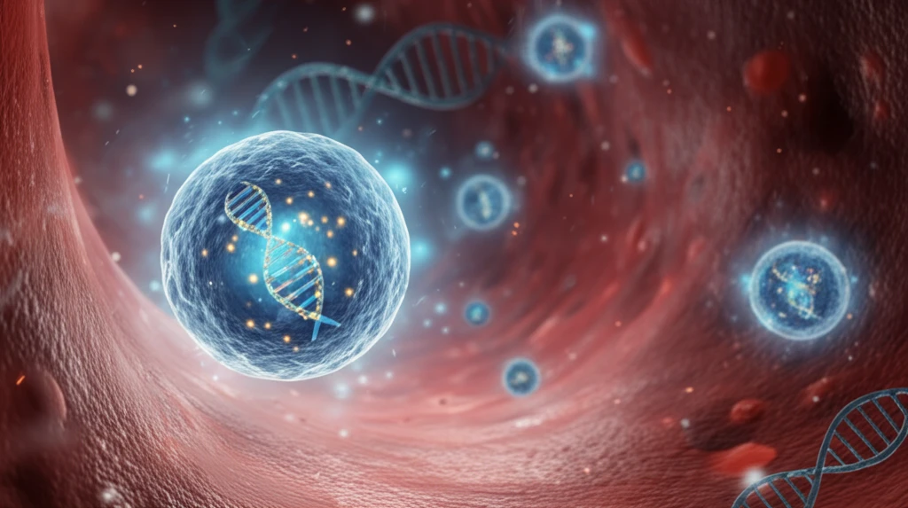
Unlock Early Disease Detection: How Extracellular Vesicles and miRNA are Changing Diagnostics

"Dive into the world of extracellular vesicles (EVs) and microRNAs (miRNAs) and discover their revolutionary role in early disease diagnosis, offering a glimpse into the future of personalized medicine."
The field of medical diagnostics is constantly evolving, driven by the need for earlier and more accurate disease detection. Among the most promising advancements are studies focused on extracellular vesicles (EVs), nanosized vesicles secreted by cells. These vesicles, including exosomes, are not merely cellular waste products; they are active messengers, carrying a diverse cargo of molecules, including microRNAs (miRNAs), that reflect the state of their parent cells.
MicroRNAs, small non-coding RNA molecules, play a crucial role in regulating gene expression and are increasingly recognized as key biomarkers for various diseases. The fact that EVs contain miRNAs derived from their cells of origin makes them particularly attractive for diagnostic purposes. Moreover, EVs are found in abundance in bodily fluids, such as blood, urine, and breast milk, making them accessible for non-invasive analysis.
Traditional methods for miRNA detection often involve complex and time-consuming procedures, including cell lysis, RNA isolation, and PCR amplification. However, recent innovations are streamlining this process, enabling simpler, faster, and more accurate detection of specific miRNAs within EVs. One such innovation is the use of molecular beacons (MBs), nanosized oligonucleotide probes that can detect miRNAs in situ, directly within the EVs, opening up new avenues for early and precise disease diagnosis.
Why Early Detection Matters
Extracellular vesicles and their microRNA cargo are being investigated as potentially useful signals for detecting disease earlier than conventional clinical changes may allow. Their appeal comes from the possibility of obtaining molecular information from accessible biofluids, although the evidence base remains developing. The diagnostic impact should therefore be viewed as promising rather than established across all diseases.
From Isolation to Clinical Use
Ultracentrifugation remains a gold-standard approach for isolating extracellular vesicles from biofluids, but its scalability and accessibility are limited. Extracellular vesicles can protect miRNAs from degradation and support delivery to specific recipient cells, yet obstacles remain before therapeutic miRNA-enriched vesicles can enter routine clinical use. Recent research also identifies analytical limitations and points to AI-driven multi-omics models as a potential way to improve clinical utility.
From Cellular Debris to Communication
Research on extracellular vesicles dates back to the 1940s, while the term “exosomes” was coined in 1981 for a major subtype of small membrane vesicles. EVs were initially understood largely as cellular waste, but research over the past two decades has increasingly recognized them as cargo-bearing structures involved in intercellular communication. Current historical reviews describe EVs as membrane-bound particles that can transport proteins, nucleic acids, lipids, and metabolites.
What are Extracellular Vesicles (EVs) and Why are They Important for Early Disease Detection?
Extracellular vesicles (EVs) are membrane-bound vesicles secreted by cells that play a crucial role in intercellular communication. They are essentially tiny packages containing proteins, lipids, and nucleic acids, including microRNAs (miRNAs). These vesicles are released by nearly all cell types and can be found in various bodily fluids, such as blood, urine, saliva, and breast milk, making them accessible for diagnostic purposes. EVs are categorized into different subtypes based on their size and biogenesis, including exosomes (30-100nm), microvesicles (100-1000nm), and apoptotic bodies (50-5000nm).
- Accessibility: EVs are readily accessible in bodily fluids, allowing for non-invasive diagnostic procedures.
- Information Richness: EVs contain a wealth of information about the cells from which they originate, providing a snapshot of cellular activity and health.
- Stability: miRNAs within EVs are protected from degradation, ensuring reliable detection and analysis.
- Disease Specificity: Certain miRNA signatures are associated with specific diseases, enabling early and accurate diagnosis.
- Early Detection: Changes in EV miRNA profiles can often be detected before clinical symptoms appear, offering the potential for early intervention.
A Broadening Research Landscape
Recent reviews are examining extracellular vesicles both as therapeutic agents and as platforms for drug delivery, with particular attention to uptake by recipient cells, signalling effects, clinical translation, and process development. EV-associated miRNA research is also being evaluated for early diagnosis in COPD, asthma, lung cancer, and COVID-19. Together, these reviews present a field that is expanding beyond basic biology while still addressing therapeutic potential and limitations.
Translation Barriers
Natural exosome applications face batch-to-batch variability, heterogeneous contents, scalability problems, and regulatory hurdles that can impede clinical translation. Circulating EV-miRNA profiles may also be affected by liver injury and dysfunction, complicating interpretation when disease-related signals overlap with organ-specific changes. These concerns show why promising biomarker findings still require careful validation and standardized characterization.
Comparing Isolation Strategies
Comparative studies are assessing how different small extracellular vesicle isolation methods affect miRNA analysis. One study compared three isolation techniques in four combinations using pooled blood plasma from four Holstein-Friesian dairy cows with similar traits and characteristics. Another comparison specifically examined plasma EVs isolated by ultracentrifugation or size-exclusion chromatography, focusing on yield, purity, and functional potential.
The Future of Diagnostics: EVs and miRNAs as Key Players
As research into extracellular vesicles and their miRNA cargo continues to advance, the potential for transforming disease diagnostics becomes increasingly clear. The development of innovative detection methods, such as molecular beacons, is streamlining the process of miRNA analysis and paving the way for more accessible and accurate early disease detection. By harnessing the power of EVs and miRNAs, we can move towards a future of personalized medicine, where diseases are detected early, treatments are tailored to the individual, and patient outcomes are significantly improved.
A Promising but Developing Field
Extracellular vesicle miRNA diagnostics offer a potentially informative way to study disease-related molecular changes in biofluids. However, the field is still developing, and differences in isolation, measurement, and validation can affect how confidently results are interpreted. The strongest path forward is likely to combine biological insight with rigorous analytical and clinical standardization.
Toward Targeted Delivery
Extracellular vesicles are emerging as biocompatible carriers for targeted drug delivery. Their future development depends on resolving persistent challenges involving production yield, cargo loading, and biodistribution. Progress in these areas will determine how broadly EV-based delivery systems can move toward practical therapeutic use.
Building Reliable Systems
The broader significance of EV and miRNA diagnostics depends on more than identifying promising molecular signals. Reliable systems will require reproducible laboratory methods, clear clinical validation, and approaches that can be implemented across different healthcare settings. Until those conditions are met, these technologies are best understood as emerging tools rather than universal replacements for established diagnostics.
From Liquid Biopsy to Patients
Effective EV isolation, enrichment, and biomarker retrieval are prerequisites for miRNA-based liquid biopsy studies, including work on non-small-cell lung cancer. In a 2025 neuroblastoma study, researchers analyzed plasma small EV miRNA from 24 patients stratified by MYCN status and risk and 10 healthy controls, then used an independent cohort of 87 patients and 47 controls for validation. Separate 2026 pancreatic cancer research reported that EV-associated miRNA signaling may contribute to immune suppression and highlighted the therapeutic potential of targeting these miRNAs to reprogram the tumor microenvironment.