PET/CT scan reveals hidden parathyroid cyst.

Parathyroid Cyst Mystery Solved: How Advanced Imaging Finds Hidden Causes of Hyperparathyroidism

"When standard tests fail, a new type of scan unveils a rare cause of elevated parathyroid hormone, leading to successful treatment."


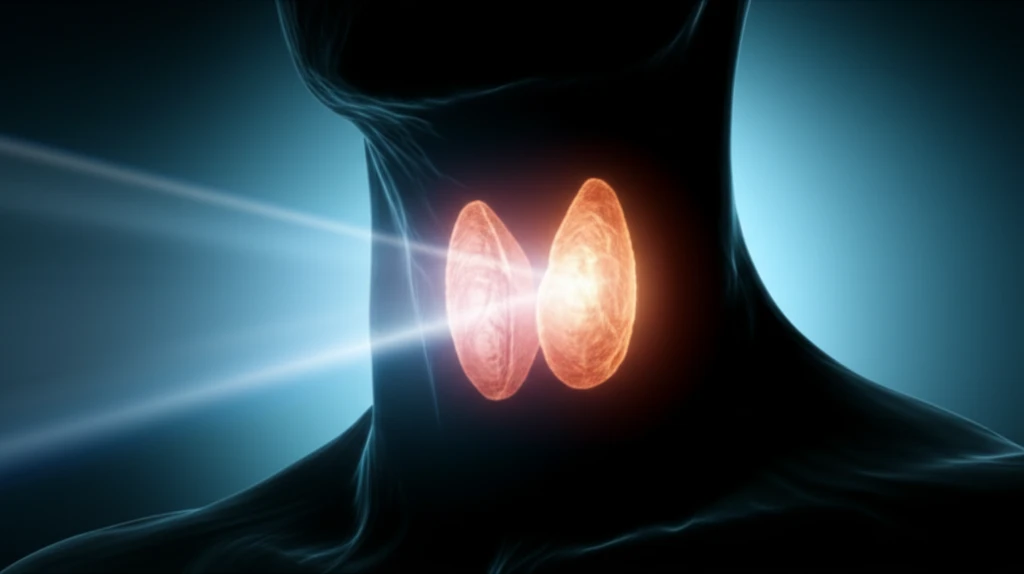
Parathyroid cysts are uncommon, fluid-filled sacs near the parathyroid glands. These cysts rarely cause problems, but sometimes they lead to primary hyperparathyroidism (PHPT), a condition where the parathyroid glands produce too much parathyroid hormone (PTH). Diagnosing these cysts can be tricky because they often don't show up on standard imaging tests.

A new imaging technique, 18F-fluorocholine positron emission tomography/computerized tomography (FC-PET/CT), is showing promise in locating these hidden cysts. This scan is usually used for cancer detection, but recent research suggests it can also pinpoint overactive parathyroid glands that are missed by traditional methods.

This article explores how FC-PET/CT successfully identified a parathyroid cyst in a woman with normocalcemic PHPT (normal calcium levels but high PTH), whose condition was previously undiagnosed. This case highlights the potential of FC-PET/CT in resolving diagnostic challenges and improving patient outcomes.

The Case: Unmasking a Hidden Cyst

PET/CT scan reveals hidden parathyroid cyst.

A 76-year-old woman with osteoporosis experienced worsening bone density despite treatment with bisphosphonates, calcium, and vitamin D. Her calcium levels were normal, but her parathyroid hormone (PTH) levels were elevated. Standard imaging, like sestaMIBI scans, failed to identify any parathyroid abnormalities, which normally indicates cause for elevated PTH.

To investigate further, doctors used an 18F-fluorocholine PET/CT scan. The scan revealed a suspicious area near her right superior parathyroid gland. This prompted surgical removal of the lesion.

  • Initial Presentation: Worsening osteoporosis despite treatment.
  • Biochemical Findings: Normal calcium, elevated PTH.
  • Imaging Results: SestaMIBI scans were negative.
  • FC-PET/CT Scan: Identified a lesion near the right superior parathyroid gland.
  • Surgical Outcome: Removal of the lesion led to resolution of hyperparathyroidism.
The removed tissue was analyzed and identified as a parathyroid cyst lined with parathyroid cells. After surgery, her PTH levels normalized, and her condition improved, demonstrating FC-PET/CT's ability to detect what other scans missed.

A New Era in Parathyroid Imaging

This case highlights the potential of FC-PET/CT in diagnosing parathyroid cysts, especially when standard imaging techniques fail. FC-PET/CT may be useful in localizing parathyroid hyperplasia and ectopic parathyroids, often missed by traditional sestaMIBI scans. Ectopic parathyroids are glands located in unusual places, making them hard to find.

While promising, more research is needed to compare FC-PET/CT directly with sestaMIBI scans in patients with PHPT. These studies will help determine the best use of FC-PET/CT in clinical practice. Comparative study will allow researchers to identify the impact of the new technology vs the current gold standard.

FC-PET/CT offers a valuable tool for pinpointing elusive parathyroid cysts, leading to timely surgical intervention and improved outcomes. This technology helps in diagnosing the right root cause and suggest treatment plan for rare diseases or conditions.

About this Article -

This article was crafted using a human-AI hybrid and collaborative approach. AI assisted our team with initial drafting, research insights, identifying key questions, and image generation. Our human editors guided topic selection, defined the angle, structured the content, ensured factual accuracy and relevance, refined the tone, and conducted thorough editing to deliver helpful, high-quality information.See our About page for more information.

Everything You Need To Know

1

What is a parathyroid cyst, and why is it important in this context?

A Parathyroid cyst is a fluid-filled sac that forms near the parathyroid glands, and they are quite uncommon. They become significant when they cause primary hyperparathyroidism (PHPT) by overproducing parathyroid hormone (PTH). The case presented involves a parathyroid cyst that led to the patient's high PTH levels, which caused worsening osteoporosis. Recognizing the parathyroid cyst's impact helps to highlight the significance of accurate diagnosis through advanced imaging.

2

What does normocalcemic hyperparathyroidism mean, and why is it relevant to this discussion?

Normocalcemic hyperparathyroidism is a condition where the levels of Parathyroid hormone (PTH) are high, but the calcium levels are normal. In this scenario, as seen in the case study, the standard imaging techniques, like sestaMIBI scans, fail to reveal any abnormalities. Consequently, the application of 18F-fluorocholine PET/CT becomes crucial to diagnose the source of increased PTH, guiding appropriate treatment for symptoms like bone loss.

3

How does 18F-fluorocholine PET/CT assist in diagnosing parathyroid issues?

18F-fluorocholine positron emission tomography/computerized tomography (FC-PET/CT) is a specialized imaging technique, commonly used for cancer detection, that can also identify parathyroid cysts. The scan proved effective in identifying a parathyroid cyst when other methods failed. This success highlights the advantage of FC-PET/CT in resolving the diagnostic challenge and improving outcomes.

4

What is the purpose of a sestaMIBI scan and why might it fail?

The sestaMIBI scan is a standard imaging technique often used to locate abnormal parathyroid glands. However, as the case study highlights, sestaMIBI scans can sometimes miss parathyroid cysts, failing to explain elevated parathyroid hormone (PTH). The failure of sestaMIBI scans in the case study emphasized the need for alternative diagnostic tools like 18F-fluorocholine PET/CT for accurate detection of the source of the patient's condition.

5

How did the surgical outcome demonstrate the value of advanced imaging?

The surgical removal of the lesion, identified as a parathyroid cyst by the 18F-fluorocholine PET/CT scan, led to the normalization of parathyroid hormone (PTH) levels and improvement in the patient's condition. The outcome demonstrates that the FC-PET/CT scan was critical for the patient's recovery. This positive outcome highlights the significance of early and accurate diagnosis, and the impact that new imaging techniques can have on patient care.

Newsletter Subscribe

Subscribe to get the latest articles and insights directly in your inbox.