
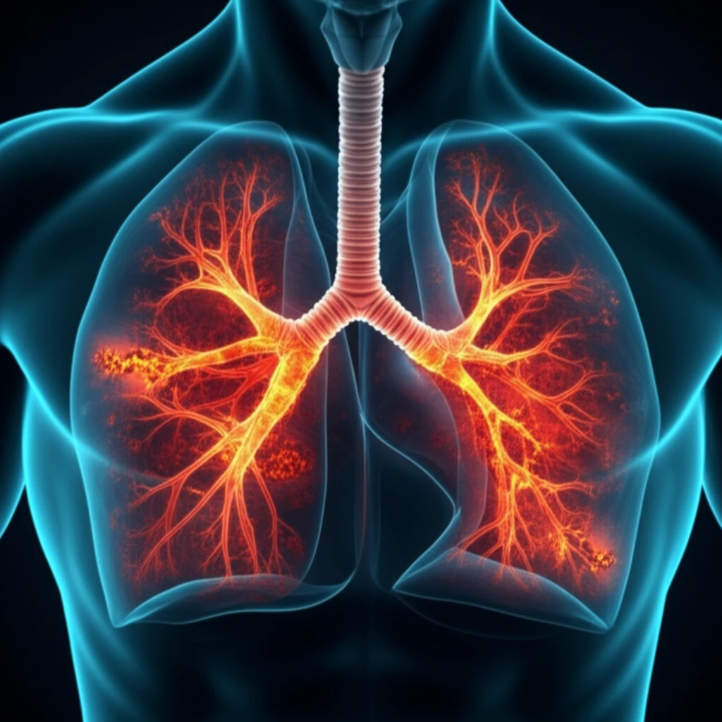
Illustration of lungs with glowing embers, symbolizing dormant TB within scar tissue.

Hidden TB? How Scans Can Spot Dormant Dangers

"Uncover the role of PET/CT scans in detecting active tuberculosis within old, healed lesions, and how it could change treatment strategies."


Tuberculosis (TB) is often thought of as a disease of the past, but it remains a significant global health concern. While active TB—the kind that makes you sick—is what most people think of, there's also such a thing as old, healed TB. This refers to lesions or scars left in the lungs after a previous TB infection. These lesions don't always mean the danger is gone; sometimes, the bacteria can linger in a dormant state, with the potential to reactivate and cause illness later.

Imagine TB as a wildfire that was seemingly put out, leaving behind charred areas. These areas, the old healed TB lesions, might still harbor embers capable of reigniting the fire. Knowing whether those embers are still glowing is crucial for preventing future outbreaks. That’s where a sophisticated tool called 18F-fluorodeoxyglucose positron emission tomography/computed tomography, or 18F-FDG PET/CT, comes into play.

A recent study delved into how PET/CT scans can help identify metabolic activity in these old TB lesions. Metabolic activity, in this case, suggests the TB bacteria might still be active, even if not causing immediate symptoms. This article explores the findings of this research, explaining how PET/CT scans work, who might benefit from them, and what the future implications could be for TB prevention and treatment.

AI Search Multiple angles on this topic

Why TB Still Matters

Tuberculosis remains a significant health concern worldwide. Its impact can vary because infection may be inactive or may progress to active illness. Understanding that distinction is important when considering how hidden infection is identified and managed.

Limits of Standard Detection

Standard TB assessment generally combines clinical evaluation with testing selected for the suspected form of infection. These approaches can be useful, but no single method provides a complete picture in every person. Symptoms, test results, and imaging may therefore need to be interpreted together.

An Airborne Disease

Tuberculosis is an infectious disease caused by bacteria, most often Mycobacterium tuberculosis. The disease primarily affects the lungs and spreads through the air, including when a person with TB coughs, sneezes, sings, or spits. Historically, TB was also known as consumption and “white death,” reflecting its long-standing impact on human health.

What is Old Healed TB and Why Does It Matter?

Illustration of lungs with glowing embers, symbolizing dormant TB within scar tissue.

Old healed TB refers to the remnants of a previous tuberculosis infection visible on a chest X-ray or CT scan. These remnants often appear as nodules (small lumps) or fibrotic scars, usually in the upper lobes of the lungs or near the hilar area (the central part of the lungs where blood vessels and airways enter). Sometimes, old healed TB can also manifest as bronchiectasis (widening of the airways) or pleural scarring (scarring of the lung lining).

The concern with old healed TB is that the seemingly inactive lesions may still contain dormant Mycobacterium tuberculosis bacteria. These bacteria can persist for years, and under the right conditions (such as a weakened immune system), they can reactivate, leading to active TB disease. Identifying individuals with old healed TB is crucial because they are at a higher risk of developing active TB in the future.

  • Increased risk of active TB: People with old healed TB are more likely to develop active TB compared to those without any signs of previous infection.
  • Potential for reactivation: Dormant bacteria can reactivate, causing new symptoms and making the person contagious.
  • Importance of monitoring: Regular check-ups and monitoring are essential to detect any signs of reactivation early.
AI Search Multiple angles on this topic

Inactive and Active TB

The Cleveland Clinic describes tuberculosis as a bacterial infection that usually affects the lungs but can spread to other organs. It reports that many people have no symptoms when they have inactive TB, while active disease may cause persistent cough, fatigue, fever, weight loss, and night sweats. TB is treatable with antibiotics, but active disease can be life-threatening if left untreated.

No Single Definitive Signal

A scan or test should not be treated as a complete answer on its own. Hidden or inactive infection may not produce obvious symptoms, while active disease can affect people differently. This means interpretation may remain uncertain without broader clinical context.

Reading Evidence Together

Different detection methods answer different questions about possible TB infection or disease. A symptom review may identify warning signs, while other assessments can provide additional evidence when symptoms are absent or unclear. Comparing results rather than relying on one signal can support a more balanced evaluation.

Think of it like a garden that once had weeds. Even after pulling the weeds, some roots might remain in the soil. If left unattended, these roots can sprout again, causing new weeds to grow. Similarly, old healed TB lesions can harbor dormant bacteria that can reactivate and cause active TB.

The Future of TB Detection: What's Next?

This research opens exciting possibilities for improving TB detection and prevention. By using PET/CT scans to identify individuals with metabolically active old healed TB lesions, doctors might be able to target preventative treatments more effectively. This could involve closer monitoring, lifestyle interventions to boost the immune system, or even targeted drug therapies to eliminate the dormant bacteria before they reactivate. Further studies are needed to fully understand the implications of these findings and to determine the best course of action for individuals with increased FDG uptake in old healed TB lesions. However, this research represents a significant step forward in our fight against TB, offering a new way to identify and manage those at risk.

AI Search Multiple angles on this topic

A Layered Assessment

The available material supports viewing TB as an infection that may be present without obvious symptoms or may develop into serious active disease. That range makes context important when clinicians assess a possible hidden danger. Scans can contribute to the evaluation, but their meaning depends on the wider clinical picture.

Earlier Recognition

Future progress will depend on recognizing TB before inactive infection or early disease becomes a larger health threat. Better integration of symptoms, testing, and imaging could make evaluation more informative. The central goal is earlier and more reliable identification without assuming that one tool can answer every question.

A Disease Beyond the Lungs

TB presents broader challenges because it spreads through the air and usually affects the lungs, while it can also involve other organs. Inactive infection may be difficult to recognize because many people have no symptoms. These features complicate detection and reinforce the need for careful public-health and clinical assessment.

Symptoms and Uncertainty

People with active TB may experience persistent cough, fatigue, fever, weight loss, or night sweats, while those with inactive TB may have no symptoms. That contrast can affect when someone seeks care or is identified for evaluation. Because active TB can be life-threatening if untreated, recognizing concerning changes and pursuing medical assessment can matter greatly.

What PET/CT findings mean in absolute terms

  • 22.37 (4.89–102.47) hazard ratioThe reported hazard ratio for culture-positive TB in the lesion group versus the normal-lung group was 22.37, with a confidence interval from 4.89 to 102.47.[1]
  • 82.6% sensitivityA meta-analysis reported FDG-PET-CT sensitivity of 82.6% for early TB detection.[2]
  • 92% sensitivity and specificityIn a macaque study before TNF neutralization, PET-measured lung inflammation together with extrapulmonary involvement predicted reactivation with 92% sensitivity and specificity.[3]

What PET activity can and cannot tell us

PET highlights glucose use by inflammatory cells, so an FDG signal reflects tissue activity rather than directly proving that TB bacteria are alive or uncontrolled.[4] [5]

The increased rate of culture-positive TB among people with subclinical lesions suggests that these scans can mark a group with elevated risk, but the wide hazard-ratio interval signals uncertainty about the size of that contrast.[1]

How PET evidence varies across settings

PET-positive lesions have also been observed within lung infiltrates and scars, and some people with these lesions developed active TB soon afterward. That pattern leaves open whether some apparent latent lesions were already early disease.[6]

PET/CT signal can vary even in macaques with culture-confirmed latent infection, and signal intensity tracks cellular glucose uptake rather than necessarily showing how well infection is controlled. Separately, two tissue-inoculation studies of Ghon foci reported sharply different estimates of MTB presence, illustrating uncertainty about bacterial persistence in such lesions.[5] [6]

Prior TB can further blur interpretation: residual changes after treatment may be difficult to distinguish from changes linked to new disease, even when the median interval since treatment was about 9 years.[7]

What PET/CT evidence still needs to resolve

  • When a PET-positive lesion appears in a scar or infiltrate, what evidence can separate stable latent infection from early active disease? Some such lesions preceded active TB, while residual changes after prior treatment can resemble new disease.[6] [7]
  • How well do risk signals observed in macaques translate to human settings beyond the specific study contexts? The available findings come from animal reactivation work and human cohort or review evidence with differing populations and measures.[3] [8]

Terms used in PET/CT evidence

FDG
FDG is a radiolabeled glucose tracer whose uptake can be located and measured by PET-CT.[6]

About this Article -

Written with AI assistance from published research, and reviewed by the Mystum team. See our About page for more information.

Everything You Need To Know

1

What exactly is Old Healed TB?

Old healed TB refers to the remnants of a previous tuberculosis infection, often visible as nodules or fibrotic scars in the lungs. These lesions result from the body's attempt to contain the Mycobacterium tuberculosis bacteria. Although they might appear inactive on imaging, these areas can still harbor dormant bacteria capable of reactivating and causing active TB.

2

Why are 18F-FDG PET/CT scans useful in detecting potential future active TB?

18F-FDG PET/CT scans are useful because they can identify metabolic activity within old healed TB lesions. This metabolic activity suggests the presence of active TB bacteria, even if the individual is not currently experiencing symptoms. The PET/CT scan uses a radioactive tracer, 18F-fluorodeoxyglucose (18F-FDG), to highlight areas of increased glucose uptake. This indicates areas with higher metabolic activity, which can be associated with the presence of active TB bacteria in the old lesions.

3

What are the implications of finding metabolically active old healed TB lesions?

Finding metabolically active old healed TB lesions using 18F-FDG PET/CT scans suggests an increased risk of future active TB disease. This means the dormant Mycobacterium tuberculosis bacteria within the lesions are potentially poised to reactivate. This information could lead to earlier interventions, such as closer monitoring, lifestyle changes, or even targeted drug therapies to eliminate the bacteria before they cause illness. The key implication is that it shifts the focus from just treating active disease to potentially preventing it.

4

Who might benefit most from PET/CT scans for detecting old healed TB?

Individuals with a history of TB, especially those with old healed lesions on chest X-rays or CT scans, could benefit from 18F-FDG PET/CT scans. High-risk groups, such as those with weakened immune systems or who have been exposed to TB, would also benefit. Those with symptoms suggestive of TB but with negative initial tests might also be considered. The scans can provide a more detailed assessment of the activity within the old healed TB lesions, potentially allowing for earlier intervention.

5

How could this new approach change the way we treat and prevent TB?

This new approach could revolutionize TB management by shifting the focus to prevention. By identifying individuals with metabolically active old healed TB lesions using 18F-FDG PET/CT scans, doctors could target preventative treatments more effectively. This might involve closer monitoring, lifestyle changes to boost the immune system, or even targeted drug therapies to eliminate the dormant Mycobacterium tuberculosis bacteria before they reactivate. The ultimate goal is to reduce the incidence of active TB and improve patient outcomes by addressing the disease at an earlier, potentially asymptomatic stage.

Newsletter Subscribe

Subscribe to get the latest articles and insights directly in your inbox.