
PET/CT Scans: A Reliable Way to Diagnose and Monitor Bone and Soft Tissue Sarcomas?

"A new analysis confirms the value of PET/CT scans in detecting and tracking sarcomas, offering hope for improved cancer care."
Osseous and soft tissue sarcomas, rare cancers affecting bone and surrounding soft tissues, often present diagnostic challenges. Accurate detection and monitoring are critical for effective treatment. While biopsies and conventional imaging techniques like X-rays, CT scans, and MRIs play a role, there's a growing need for more precise diagnostic tools.
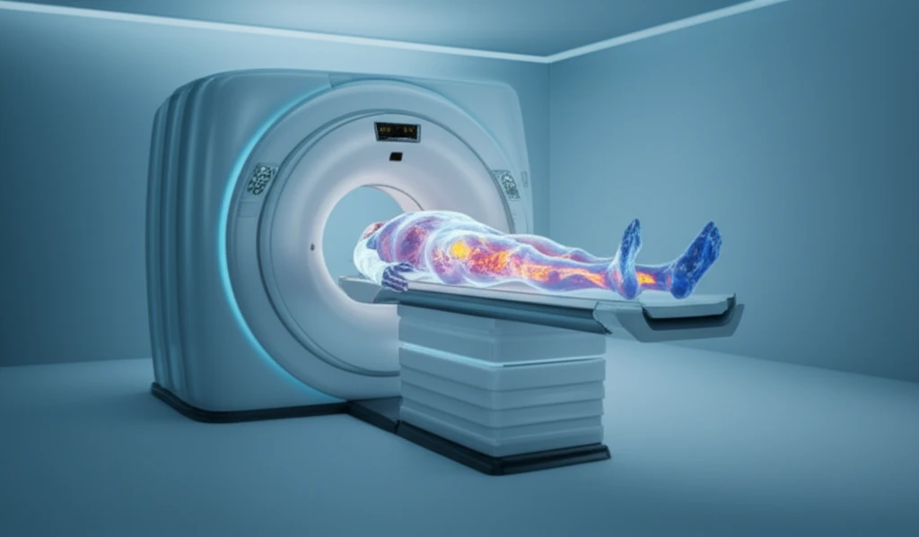
Positron Emission Tomography (PET) scans, combined with Computed Tomography (CT) in a PET/CT scan, offer a promising solution. PET scans measure the metabolic activity of cells, while CT provides detailed anatomical images. This combination allows doctors to visualize both the structure and function of tissues, potentially differentiating between benign and malignant tumors.
A recent study analyzed existing research to determine how effective PET/CT scans are in diagnosing and monitoring sarcomas. This article breaks down the findings of this analysis, explaining the potential benefits and limitations of using PET/CT scans in sarcoma management.
Diagnosis Relies on Imaging and Tissue Examination
Soft tissue sarcoma diagnosis typically starts with imaging of a suspicious lump and proceeds to a tissue biopsy examined under a microscope, sometimes supplemented by molecular testing; no single scan or blood test confirms the diagnosis on its own (Science Insights). The role of 18F-FDG PET/CT in the initial diagnostic work-up of bone and soft tissue tumors is not well established, and the cited review says it is not recommended for the initial local imaging work-up or for diagnosing sarcoma (ScienceDirect). Separately, imaging recommendations call for chest CT in all patients and regional CT or MRI to assess lymph-node metastases in certain high-grade malignancies, including angiosarcoma, synovial sarcoma, and epithelioid sarcoma (Clinical Radiology).
The Accuracy of PET/CT Scans in Diagnosing Sarcomas
The study reviewed data from multiple published articles, focusing on the ability of PET/CT scans to accurately identify sarcomas. The analysis included a significant number of patients and lesions, providing a robust dataset for evaluation.
- High Sensitivity: PET/CT scans demonstrated a high sensitivity, meaning they were good at correctly identifying cancerous lesions. Specifically, the sensitivity was around 90%, indicating that the scan would correctly identify 90 out of 100 sarcomas.
- Good Specificity: The scans also showed good specificity, meaning they were able to accurately distinguish between cancerous and non-cancerous tissues. The specificity was approximately 89%, indicating that the scan would correctly identify 89 out of 100 benign lesions.
- Combined Results: When both cancerous and non-cancerous tissues were examined with lesion-based analysis the accuracy was about 96% for the scans.
Research Spans Diagnostic Accuracy and Treatment Monitoring
A 2024 bibliometric analysis examined publications from the preceding 13 years on PET/CT use in diagnosing and treating bone and soft tissue sarcomas (Frontiers in Oncology). In a single-center study of 255 Chinese patients with primary musculoskeletal tumors, SUVmax-based diagnosis had reported sensitivities of 96.6% for primary malignant bone sarcomas and 91.2% for soft tissue sarcomas (British Journal of Radiology). A separate report found that combining 18F-FDG PET metabolic data with MRI improved the reliability and efficiency of planning and monitoring isolated limb perfusion treatment for soft tissue sarcoma (Oncology). A 2025 review describes 18F-FDG PET/CT as a modality for sarcoma diagnosis, staging, and treatment monitoring that complements conventional imaging (Molecular Imaging and Biology).
Potential Value Comes With Practical Limitations
One article says PET/CT’s high sensitivity and specificity may help clinicians assess whether a suspicious mass is sarcoma and support early diagnosis and treatment planning (MyStum). However, a management review identifies radiation exposure, false-positive findings caused by post-treatment inflammation, and infrastructure requirements as significant limitations of PET/CT (AIIM Research). The review also reports that PET/CT has contributed to standardized biobanks combining imaging, genomic, and clinical data, which it says may support AI-driven predictive models for individualized sarcoma treatment.
The Future of Sarcoma Diagnosis with PET/CT
This research strengthens the evidence supporting the use of PET/CT scans in the diagnosis and management of bone and soft tissue sarcomas. The ability to accurately detect and monitor these tumors can lead to earlier diagnosis, more effective treatment plans, and ultimately, improved outcomes for patients.
Assessing PET/CT’s Effect on Treatment Decisions
In a study evaluating the impact of 18F-FDG PET/CT on patient management and treatment, physicians from the institution’s multidisciplinary sarcoma team first indicated their preferred therapeutic strategy using patients’ clinical data and available diagnostic imaging without knowing the PET/CT findings. This setup provided a basis for evaluating the scan’s impact on treatment decisions (PMC).
Case Reports Illustrate Multimodal Assessment
A 2026 case report describes a diagnostic work-up combining ultrasound, MRI, CT, and whole-body 18F-FDG PET/CT to evaluate tumor extent, possible bone involvement, and regional lymph-node disease (Frontiers in Nuclear Medicine). A separate Frontiers case report focuses on CT characteristics and the pathological basis for diagnosing primary liver carcinosarcoma, also referred to in the source snippet as primary hepatocellular carcinoma sarcoma. A retrospective study of 89 patients examined the diagnostic and prognostic value of 18F-FDG PET/CT in the initial assessment of high-grade bone and soft tissue sarcoma (Journal of Surgical Oncology).
While promising, it's important to acknowledge some limitations. The analysis included a relatively small number of patients in some studies. Additionally, the high cost and limited availability of PET/CT scans may restrict their widespread use.
Despite these limitations, PET/CT scans hold significant potential for transforming sarcoma care. As technology advances and costs potentially decrease, PET/CT imaging may become an increasingly integral tool for diagnosing and monitoring these challenging cancers.