
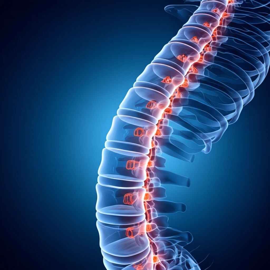
Neck Stability: How Atlantoaxial Fusion Can Help

"A Comprehensive Guide to Atlantoaxial Instability and the Role of Fusion Techniques in Restoring Neck Stability"
The upper cervical spine, comprising the atlas (C1) and axis (C2) vertebrae, is a marvel of engineering, designed for flexibility and support. Unlike the more uniform lower cervical vertebrae, C1 and C2 boast unique anatomical features that allow for a greater range of motion, particularly rotation. This specialized structure, however, comes at a cost: increased vulnerability to instability.
Atlantoaxial instability (AAI) occurs when the ligaments and bony structures that stabilize the C1-C2 joint are compromised. This compromise can lead to excessive movement, pain, and even neurological deficits. Understanding the causes, symptoms, and treatment options for AAI is crucial for those experiencing neck pain, stiffness, or neurological symptoms.
This article delves into the complexities of AAI, exploring the various fusion techniques used to restore stability and improve patient outcomes. It's inspired by a recent study examining the effectiveness of different fusion methods, including posterior wiring, transarticular screw fixation, and posterior screw-rod fixation.
A Common Instability with Varying Severity
Atlantoaxial instability is reported as the most common cervical instability seen in patients with rheumatoid arthritis, though its course can differ between patients, with varying degrees of severity and symptoms. There is very limited data on atlantoaxial instability in patients with psoriatic arthritis, an area researchers note needs further study. In children, the excess mobility of the atlantoaxial joint can, in severe cases, cause the bones of the neck to compress the spinal cord, leading to temporary or permanent spinal cord injury. The joint's normal role is to allow the neck to rotate and extend, which is why instability at this level carries such significant risk.
A Transitional Zone Prone to Failure
The atlantoaxial joint is described as an important transitional zone in the cervical spine, prone to instability through both degenerative and traumatic processes. Adult causes of atlantoaxial instability include conditions such as Down syndrome and rheumatoid arthritis, and the instability reflects excessive movement between the atlas (C1) and axis (C2) vertebrae that can compress the spinal cord or nerve roots. Because conventional fixation has limitations, surgeons have explored alternatives such as the Kishigami atlantoaxial tension band, which remains in the epidural space. A related study developed a modified dorsal wiring method based on that approach and evaluated it in toy breed dogs with atlantoaxial instability.
A Condition Slow to Be Recognized
Early manifestations of atlantoaxial instability are often subtle and can be difficult to elicit, particularly in individuals with intellectual disability, so diagnosis may be delayed and clinical suspicion often rests on indirect or inferred symptoms. Because the presentation can be vague, awareness of the condition has at times grown through individual case experiences rather than systematic study. One widely discussed report describes severe dizziness and vomiting after massage therapy that clinicians attributed to atlantoaxial instability or vertebrobasilar insufficiency, with lessons for health care consumers. Such accounts helped illustrate how easily the condition can be missed in everyday care.
Understanding Atlantoaxial Instability
Atlantoaxial instability arises from a variety of factors that compromise the structural integrity of the C1-C2 joint. These can be broadly categorized as traumatic, congenital, and acquired conditions. Traumatic injuries, such as fractures of the odontoid process (dens) or rupture of the transverse ligament, are common causes. Congenital conditions like Os odontoideum, where the odontoid process fails to fully develop, can also lead to AAI. Acquired conditions such as rheumatoid arthritis can erode the ligaments and bony structures, resulting in instability.
- A thorough neurological assessment helps identify any nerve involvement.
- Dynamic X-rays, which are taken while the patient flexes and extends their neck, can reveal abnormal movement at the C1-C2 joint.
- MRI scans provide detailed images of the ligaments, spinal cord, and surrounding tissues, helping to identify the cause and extent of the instability.
- CT scans are for bony structures that provides detailed images of fracture.
Multifactorial Causes, Evolving Evidence
Recent literature reviews characterize atlantoaxial instability as abnormal motion between the atlas (C1) and axis (C2) that can lead to spinal cord compression and significant neurological deficits, with a multifactorial etiology. Newer research also shows that instability can arise as a complication of traction therapy, and investigators have sought to identify risk factors for atlantoaxial instability during traction and to determine whether the instability reverses once traction ends. In the related area of craniocervical instability, researchers note that very little research has explored the many problems seen in patients with hypermobile Ehlers-Danlos syndrome, and the diagnostic criteria and surgical indications are not universally agreed upon. This means that for connective tissue populations, both diagnosis and treatment decisions remain matters of ongoing debate.
Debates Over Risk and Screening
Atlantoaxial instability is defined as excessive mobility at the articulation between the atlas (C1) and axis (C2), which can potentially result in spinal cord compression. This risk has drawn longstanding attention: in 1984, the Committee on Sports Medicine of the American Academy of Pediatrics published in this journal a statement on the 'remarkably high' incidence of atlantoaxial instability among individuals with Down syndrome. A more recent analysis in the same journal offers 'a fresh look' at the condition, reflecting ongoing reassessment of earlier claims about its frequency and significance. Because the diagnosis itself rests on findings of excessive mobility, what counts as clinically meaningful instability remains open to interpretation.
Comparing Fixation Techniques
Biomechanical studies have compared anterior and posterior stabilization methods for atlantoaxial instability, evaluating the stability provided by pedicle screws against various established posterior atlantoaxial fixations. In one such comparison, the anterior transarticular crossing screw was reported to be a biomechanically effective alternative or supplemental method, providing basic evidence for clinical application. Clinical comparisons have likewise examined two surgical options, C1-C2 transarticular screw fixation versus C1 lateral mass plus C2 pedicle screw fixation, for treating atlantoaxial instability. Additional clinical experience with an alternative C-1 screw placement technique, used in 26 patients across three institutions by three surgeons, has also been reported using standard lateral fluoroscopy and fully threaded polyaxial screws.
The Road to Recovery
Atlantoaxial fusion is a powerful tool for restoring stability and improving the quality of life for individuals with AAI. While the recovery process requires commitment and patience, the long-term benefits of a stable and pain-free neck are well worth the effort. By understanding the causes, symptoms, and treatment options for AAI, you can take control of your health and embark on the path to recovery.
Instability as a Surgical Variable
Experts have analyzed the role of atlantoaxial instability in the management of ossification of the posterior longitudinal ligament (OPLL) that extends into the high cervical region, above the lower border of the C3 vertebra. In this context, atlantoaxial instability is examined as a factor that may influence how high cervical OPLL should be approached and treated. The series that informed the analysis was made up entirely of male patients, a composition noted when discussing the findings. This work highlights how recognizing instability at the C1-C2 level can be central to surgical decision-making in complex cervical pathology.
Surgical Advances and Recovering Lives
As understanding of atlantoaxial instability grows, resources aimed at families emphasize practical information on causes, symptoms, and treatment options, and on how to support children living with the condition. Surgical outcomes have also become more encouraging: after surgery to correct atlantoaxial instability, one child described at Boston Children's Hospital was able to return home within a few weeks, and the rehabilitation nurses and therapists who knew her were 'amazed' by her progress. Such recovery stories point to the potential for surgical correction to substantially change a child's trajectory. The broader frontier lies in translating these individual successes into clearer guidance on diagnosis and the timing of treatment.
Access and Awareness Gaps
Because atlantoaxial instability can present subtly and its diagnosis often depends on specialized imaging and clinical expertise, access to care is likely uneven across communities. Patients may face long delays before the condition is recognized, and not every center offers the surgical expertise needed for C1-C2 reconstruction. Awareness among general practitioners and the public, alongside consistent referral pathways, remains an important challenge. The systemic issues are therefore as much about recognition and access as they are about the surgical technique itself.
Recognition Can Change Outcomes
For many patients, atlantoaxial instability occurs alongside cervical spondylotic disease at a single or multiple spinal levels, and the association between the two conditions has been specifically assessed in the literature. Identifying atlantoaxial instability in such patients matters because its presence may change how treatment is planned, and the implications of recognizing and treating the instability in this association have been highlighted. The human impact is direct: instability at the atlantoaxial joint can threaten spinal cord function, so detecting it can alter a person's neurological future. This is why clinical attention to the C1-C2 segment, even when more obvious cervical problems are present, can be life-changing for individual patients.