
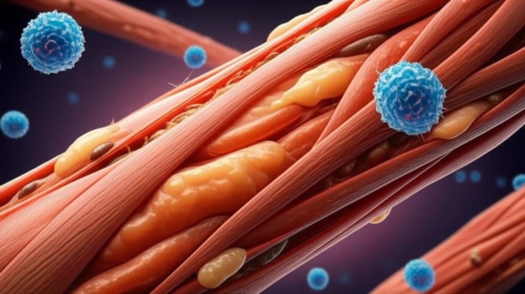
Muscle Macrophages: How These Immune Cells Impact Muscle Growth and Repair

"Discover the surprising role of macrophages, a type of immune cell, in muscle development and how they could hold the key to better treatments for muscle-related conditions."
For years, we've viewed muscle tissue primarily through the lens of exercise and nutrition, focusing on protein intake and workout routines to build strength and endurance. But what if there's another key player in muscle health that we've largely overlooked? Recent studies are shedding light on the critical role of immune cells, specifically macrophages, in muscle development and repair. These cells, traditionally known for their role in fighting off infections, are now being recognized for their surprising impact on muscle tissue.
Intramuscular fat, the kind that nestles within our muscles, isn't just about aesthetics; it's a real concern for several health issues like muscular dystrophies, metabolic disorders, and even the effects of aging. Scientists are really zoning in on these special cells called fibro-adipogenic progenitors (FAPs). Think of them as the main source for creating those unwanted fat cells inside our muscles. However, we are still figuring out the best ways to control how these FAPs behave, especially in us humans. In this study, the researchers wanted to understand if the macrophages can also control human FAP adipogenesis.
A groundbreaking study published in Scientific Reports has uncovered the complex relationship between different types of macrophages and fibro-adipogenic progenitors (FAPs), the cells responsible for creating intramuscular fat. The study reveals that certain macrophages can either promote or inhibit the creation of fat cells within muscles, offering new insights into potential treatments for muscle-related conditions. This is a game changer because understanding these interactions could lead to more effective therapies for muscle disorders.
A Developing Field
Research increasingly indicates that muscle macrophages influence inflammation, tissue cleanup, and repair after muscle injury. Their effects may depend on timing, local signals, and the transition between inflammatory and reparative states. However, the field remains too context-dependent to support a single universal estimate of their impact on muscle growth or recovery.
Macrophages in Muscle Repair
The standard model describes macrophages as shifting from pro-inflammatory, M1-like activity toward reparative, M2-like activity during skeletal muscle regeneration. These cells help clear debris, regulate inflammation, activate satellite cells, and support tissue remodeling. Studies report that macrophage depletion impairs muscle regeneration, while mechano growth factor may partly reduce that impairment. Research also indicates that glutamine availability and macrophage glycolytic or oxidative metabolism can influence muscle stem-cell activation, showing why a simple M1-versus-M2 framework may not capture the full process.
From Inflammation to Regeneration
Early and continuing research has expanded the view of skeletal muscle macrophages from general inflammatory cells to diverse participants in muscle homeostasis, injury repair, and muscular dystrophy. Reviews now emphasize their roles in inflammation, phagocytosis, stem-cell activation, and tissue repair. At the same time, researchers acknowledge that the origins, heterogeneity, and activation mechanisms of these cells remain incompletely understood.
Macrophages: Muscle's Unlikely Allies or Silent Saboteurs?
Macrophages, a type of white blood cell, are like the cleanup crew and security guards of our bodies. They engulf and digest cellular debris, foreign substances, microbes, and cancer cells. It's only recently that scientists have started to understand the varied roles they play in muscle tissue.
New Cellular and Metabolic Insights
A 2025 Nature Communications study identified monocytes/macrophages and fibro-adipogenic progenitors as pivotal players in human muscle regeneration through single-cell and spatial deconvolution analysis. Recent reviews also describe pathogenic macrophage populations with altered metabolic and epigenetic profiles in chronic inflammatory muscle diseases, including muscular dystrophies. A separate review highlights a dual role: macrophages can remove necrotic tissue and release factors such as IGF-1 and IL-10 that support satellite-cell activation, yet macrophage activity can also contribute to muscle pathology.
Limits of the Evidence
Macrophage research does not yet establish that increasing macrophage activity will consistently improve muscle growth or repair. Effects may vary with disease state, timing, metabolic conditions, and the specific macrophage population involved. Findings from animal models, cellular studies, and human research may therefore not translate directly into a single treatment strategy.
Context Determines the Outcome
Macrophages can support regeneration in one setting while contributing to persistent inflammation, fibrosis, or muscle loss in another. Comparisons across studies should account for differences in injury models, disease context, timing, and methods used to identify macrophage states. The available evidence supports a context-dependent role rather than a uniformly beneficial or harmful effect.
Looking Ahead: Macrophages as Therapeutic Targets
These findings open up new possibilities for treating muscle-related conditions characterized by excess fat accumulation. By understanding how to manipulate macrophage behavior, scientists may be able to develop therapies that promote muscle regeneration and reduce intramuscular fat. This could be particularly beneficial for individuals with muscular dystrophies, metabolic disorders, and age-related muscle wasting. These results may lead to the creation of innovative regenerative medical techniques to stop fat accumulation in diseased muscles linked to chronic inflammation.
A Coordinated Repair System
Current evidence portrays muscle macrophages as adaptable coordinators whose effects depend on cellular identity, timing, and metabolism. Temporal single-cell sequencing identified a macrophage subset with high Gpnmb expression that also expressed factors involved in muscle regeneration, including Igf1. Other research links macrophage glutamine secretion and broader glycolytic and oxidative metabolism to muscle stem-cell activation. Reviews have also examined their possible contribution to skeletal muscle hypertrophy in both animal and human experiments, although that contribution remains an active research question.
Reprogramming Macrophage Metabolism
A major future direction is to understand whether macrophage metabolism can be deliberately reprogrammed to improve muscle repair. Emerging research is examining metabolic pathways, communication between macrophages and other cells, and therapeutic strategies centered on macrophage metabolic states. The central challenge is translating these insights into safe and effective interventions for muscle-wasting conditions. Such approaches will need to account for the possibility that the same immune programs may help regeneration in one context but worsen chronic disease in another.
Repair Within a Larger System
Muscle macrophage behavior is shaped by the surrounding tissue and broader physiological environment. Injury, aging, chronic inflammation, and muscle-wasting conditions may alter the signals that guide macrophage activity. This makes muscle repair a systems problem rather than an isolated immune-cell process. Research will need to connect local macrophage biology with the wider factors affecting tissue health.
What This Means for Recovery
For people recovering from muscle injury or living with muscle-wasting conditions, macrophage research offers a possible explanation for why repair does not proceed identically in every situation. The findings suggest that immune-cell timing and state may matter alongside conventional approaches to recovery. However, the research does not yet justify treating macrophages as a simple target for boosting muscle growth. Human applications will require evidence that interventions improve meaningful recovery outcomes safely.