
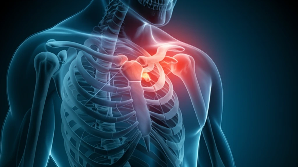
Decoding Shoulder Pain: Understanding Shoulder Dislocation and Acromioclavicular Joint Injury

"Navigating the complexities of shoulder injuries: A closer look at the rare combination of shoulder dislocation and acromioclavicular joint injury, its diagnosis, and treatment options."
Shoulder injuries can significantly impact your daily life, from limiting your ability to reach for a cup of coffee to hindering your performance in sports. While some shoulder problems are relatively common, others are exceedingly rare. This article delves into one such unusual occurrence: the simultaneous dislocation of the shoulder joint (glenohumeral joint) and injury to the acromioclavicular (AC) joint.
The shoulder is a complex structure composed of several joints, ligaments, and muscles working together to provide a wide range of motion. The glenohumeral joint, often simply called the shoulder joint, is a ball-and-socket joint where the head of the humerus (upper arm bone) fits into the glenoid fossa of the scapula (shoulder blade). The AC joint, on the other hand, is where the clavicle (collarbone) meets the acromion, a bony projection of the scapula.
Understanding the intricacies of these joints and the potential injuries that can occur is crucial for effective diagnosis and treatment. In the following sections, we'll explore the causes, symptoms, diagnostic methods, and treatment options for the rare combination of shoulder dislocation and AC joint injury, providing you with a comprehensive guide to navigate this complex condition.
A Common Injury With High Recurrence Risk
Shoulder dislocation is one of the most frequently seen joint injuries, and the most common direction of displacement is anterior, with the arm bone slipping forward and down out of the joint. A Greek study of 308 patients (170 men and 138 women) found that the most frequent mechanism of injury was falling, and that 92% of reductions took place in the emergency department. The single most important risk factor for recurrence is the age at which the first dislocation occurs: in patients under 20, the chance that the shoulder will come out again after a first dislocation can exceed 70%, according to one orthopedic practice. Large pooled analyses continue to study these outcomes; a recent review of 34 studies covering 2,222 shoulder dislocations examined randomized trials that compared immobilization with arthroscopic Bankart repair after a first dislocation.
Closed Reduction and Its Cautions
Anterior dislocations account for approximately 95-98% of all shoulder dislocations and are the most common type seen in urgent care and emergency medicine settings, usually arising from forced abduction and external rotation of the arm. The standard acute treatment is closed reduction in the emergency department, followed by a period of immobilization and rehabilitation. However, this approach has clear limitations: for posterior dislocations, standard closed reduction is contraindicated in cases of delayed presentation of more than 3-6 weeks or when a large humeral head defect is present. Recognition itself is a challenge, as posterior dislocations are notoriously easy to miss and the diagnostic approach for anterior versus posterior injuries differs significantly. For dislocations without severe fractures or complications, non-surgical management is generally recommended to restore normal function and stability.
From Ancient Egypt to the Modern Clinic
Shoulder dislocation is one of the oldest recognized injuries in medicine, documented in Egyptian tomb murals as early as 3000 BC, where a manipulation resembling the Kocher technique is depicted. Historically it has been regarded as the most common large-joint dislocation, occurring in anterior, posterior, and inferior directions, most often anteriorly. Contemporary data still reflect this prominence: shoulder dislocations account for 50% of all major joint dislocations. The typical mechanism recorded in modern training resources, forced abduction with external rotation driving the humeral head between the glenoid and coracoid process, mirrors the injury patterns described over centuries of clinical observation.
Understanding the Rare Combination: Glenohumeral Dislocation and Acromioclavicular Disjunction
The simultaneous occurrence of glenohumeral dislocation and acromioclavicular disjunction is an exceptionally rare phenomenon. Medical literature documents only a handful of such cases, highlighting the unusual nature of this combined injury. These injuries typically result from high-impact trauma, such as a fall directly onto the shoulder. The force of the impact causes damage to the ligaments and supporting structures of both the glenohumeral and AC joints.
- High-impact trauma: Falls or direct blows to the shoulder.
- Glenohumeral joint dislocation: Displacement of the humerus from the glenoid fossa.
- AC joint injury: Sprains, subluxations, or dislocations of the acromioclavicular joint.
- Ligament damage: Tearing or stretching of ligaments in both joints.
A Field Defined by Ongoing Review
Research into shoulder dislocation continues to build on centuries of clinical practice, and current reviews note that the reduction methods described in the Hippocrates Corpus have been repeatedly described and adjusted by later physicians. A literature review published in the August 2012 issue of the Journal of the American Medical Association also surveyed the evidence on shoulder joint dislocation, underscoring that it remains among the most studied joint injuries. Ongoing coverage of dislocation research spans everything from acute management refinements to long-term outcomes, and news aggregators routinely track newly published studies in the field. The result is a body of literature that keeps revisiting the same central question: how to reduce recurrence rates after a first dislocation.
Treatment Pitfalls and Safer Techniques
Despite a well-established standard approach, shoulder dislocation care is prone to recurring failures, and clinicians commonly cite five treatment mistakes that compromise outcomes, from improper technique to inadequate follow-up. The problem of recurrence is real: once a shoulder has been dislocated, the likelihood of it happening again can increase, especially without proper treatment and rehabilitation. In response, some teams have argued that traditional reduction methods can be improved upon; a study of the Davos technique concluded it is easy, non-traumatic, very well tolerated, and most of all safe, being complication-free and giving reproducible results comparable or superior to other reduction techniques. These critiques push the field toward gentler, more reliable methods of restoring the joint.
Dislocation Versus Separation: Sorting Out the Differences
Shoulder dislocations are classified into two main categories by the direction the humerus travels: in an anterior dislocation the upper arm bone is forced out of the front of the shoulder socket, while a posterior dislocation involves the bone moving out of the back of the socket. The key difference between the two always comes down to which direction the humeral head moves relative to the socket. A separate distinction is often confused with dislocation: a shoulder separation, or AC joint injury, involves the joint where the collarbone meets the shoulder blade rather than the ball-and-socket glenohumeral joint itself. The two conditions share similar symptoms, which is why clinicians emphasize careful comparison, both between anterior and posterior dislocations and between a true dislocation and an AC joint separation.
Treatment and Rehabilitation: Steps to Recovery
The treatment approach for combined glenohumeral dislocation and AC joint injury depends on the severity of the injuries and the individual's specific needs. Initially, the glenohumeral dislocation must be reduced, which involves manually manipulating the humerus back into its normal position within the glenoid fossa. This is typically performed under anesthesia to minimize pain and muscle spasm. Once the shoulder is reduced, the focus shifts to stabilizing both the glenohumeral and AC joints. In some cases, non-operative management, such as immobilization in a sling, pain management, and physical therapy, may be sufficient. However, more severe AC joint dislocations or persistent glenohumeral instability may require surgical intervention to repair or reconstruct the damaged ligaments. Rehabilitation plays a vital role in restoring shoulder function after either non-operative or surgical treatment. Physical therapy programs focus on regaining range of motion, strengthening the surrounding muscles, and improving proprioception (the body's awareness of its position in space). With appropriate management and diligent rehabilitation, most individuals can achieve a satisfactory recovery and return to their desired activities.
Expert Views on First-Time Dislocations
Synthesizing the evidence, experts agree that forward (anterior) dislocation is by far the most common pattern, occurring when the upper arm bone is displaced from the cup-shaped socket of the shoulder blade, though the shoulder can also slip backward or downward. Posterior instability and dislocations are less common than anterior injuries but are much more frequently missed, and diagnosis is made radiographically in the setting of acute dislocations. On management, opinion leaders such as Eiji Itoi, MD, PhD, an emeritus professor of Tohoku University in Japan, have published editorials laying out their personal approach to the first-time shoulder dislocation, reflecting genuine disagreement in the field about how aggressively early surgery should be pursued. Institutions like the Acibadem Healthcare Group have responded with comprehensive treatment protocols that emphasize patient well-being and long-term shoulder stability.
Reducing Recurrence: The Next Frontier
The future of shoulder dislocation care is focused on prevention of recurrence, and systematic reviews confirm that acute treatment centers on closed reduction, which should be performed as soon as possible either on the field or in an emergency department. Yet even with prompt care, some patients develop recurrent dislocations or symptomatic subluxations during daily activities, which remains the core unsolved problem. The shoulder joint is especially prone to dislocation precisely because of its extraordinary range of motion, so future protocols aim to pair rapid reduction with rehabilitation designed to protect that mobility without sacrificing stability. Public education also plays a role, since experts stress that people should not try to pop the joint back in themselves and should instead seek proper medical care after accidents, falls, or sports injuries.
Care Access, Neglect, and Holistic Recovery
While anterior shoulder dislocation is a disabling injury affecting all age groups, access to adequate treatment is far from universal, and neglected dislocations occur in unconscious patients or as a result of inadequate treatment, which is common in low-income countries. These gaps in care create long-term problems such as the Hill-Sachs lesion that complicate later surgical management. Where treatment is available, practitioners increasingly take a holistic view, considering not only the physical injury but its broader impact on a patient's movement, confidence, and function. Rare and unusual cases also demand attention, such as the extremely uncommon dislocation of a shoulder at birth, which results from traumatic birth injury, brachial plexus injury, or a true congenital dislocation developed in utero with no associated injury.
Lives Affected: Athletes, Patients, and Recovery
Research into first-time traumatic anterior shoulder dislocation shows it negatively impacts quality of life, shoulder pain, and function, and although these measures improve over time, people with recurrent shoulder instability report poorer quality of life even 12 months after the initial injury. The personal toll is visible in cases like that of Olympic boxer Anthony Ogogo, whose dislocated shoulder was extremely painful and required hospital treatment to restore the joint's structure, with ibuprofen and cold compresses used for relief. Rare variants add another layer of impact, with inferior dislocations accounting for roughly 0.5% of cases as the rarest form of the injury. In emergency settings, bedside tools are evolving too, with case-control studies examining ultrasound as a way to verify reduction before radiography.