
Decoding Calmodulin: How Understanding This Protein Could Unlock Better Health

"Researchers map the conformational landscape of calmodulin, revealing new insights into its function and potential therapeutic applications."
In the intricate dance of cellular life, proteins play the lead roles, orchestrating everything from muscle contraction to nerve signaling. Among these molecular maestros, calmodulin stands out as a versatile calcium-binding protein. It acts as a critical messenger, relaying calcium signals to trigger a cascade of downstream events. New research sheds light on calmodulin’s structural dynamics, potentially paving the way for innovative therapies.
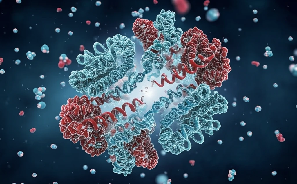
Calmodulin's structure is unique, featuring two EF-lobes (N-lobe and C-lobe) that respond differently to calcium. Think of these lobes as specialized hands, each with its own grip and set of functions. While they share similarities, their subtle differences are key to calmodulin's diverse roles. Understanding these nuances is crucial for designing targeted interventions that harness calmodulin's power.
Imagine trying to understand a complex machine without a blueprint. That's the challenge scientists face when studying proteins. But what if you could map out every possible movement and interaction? Recent work has done just that for calmodulin, creating a 'conformational landscape' that reveals how its structure changes in response to calcium and other signals. This landscape is more than just a pretty picture; it's a roadmap for developing new drugs and therapies.
Calmodulin's Central Role in Cellular Regulation
Calmodulin is a heat-stable, low-molecular-weight, multifunctional protein found in all eukaryotic cells, present especially in brain and heart tissue. It acts as a key intracellular calcium receptor: when calcium ions bind, calmodulin undergoes conformational transitions that allow it to specifically activate a large number of target proteins and regulate diverse cellular processes. Despite being identified decades ago, calmodulin continues to be studied as one of the most versatile signalling mediators in biology. Its ubiquity across eukaryotic organisms underscores its fundamental importance to life.
Methods for Studying Calmodulin and Their Challenges
Researchers have employed multiple methods to isolate and characterize calmodulin. Traditional approaches include purifying calmodulin to homogeneity from bovine brain using fluphenazine-affinity chromatography, and quantifying it via radioimmunoassay. Amino acid analysis has been used to determine the concentration of standard calmodulin preparations, though discrepancies between amino acid analysis and immunoassay results have been noted, suggesting methodological limitations. More recently, chemoenzymatically modified calmodulin combined with click chemistry has been developed to streamline affinity purification conjugation to Sepharose resin, replacing a previously multi-step process. Liquid chromatography-tandem mass spectrometry (LC-MS/MS) approaches have enabled estimation of lysine and arginine methylation stoichiometries for calmodulin, though limitations of amino acid analysis have hampered broader general application of these techniques.
Defining Calmodulin: A Foundational Protein
Calmodulin is defined as a calcium-binding protein that regulates cellular processes. Its discovery established it as a pivotal intermediary in calcium signalling — a second-messenger system now recognized as fundamental to virtually all eukaryotic cell biology. The recognition of calmodulin as a distinct regulatory protein, rather than merely one of many calcium-binding species, was itself a milestone that redirected research toward understanding calcium-dependent signal transduction pathways. Unfortunately, the source material available for this subsection provides only this high-level definitional context, with detailed historical milestones lying beyond the scope of the listed references.
Mapping Calmodulin's Landscape: A New Perspective
The recent study by Kawasaki and Kretsinger delves into the conformational landscape of calmodulin, focusing on the differences between its N- and C-lobes. By analyzing the helix positions within these lobes, the researchers have created a detailed map of how calmodulin's structure shifts and changes. This approach uses a pseudo-two fold axis for alignment, allowing for a precise comparison of the lobes' structural dynamics.
- Calcium binding: Determines how calmodulin interacts with target proteins.
- Hydrophobic interactions: Crucial for calmodulin's stability and function.
- Structural changes: Influence calmodulin's activity and interactions.
- EF-hand dynamics: Dictate how calmodulin responds to calcium signals.
Advancing Calmodulin Research with Modern Techniques
Current calmodulin research leverages advanced tools to probe the protein's function. A project using the Canadian Light Source synchrotron facility aims to deepen understanding of calmodulin's role in regulating heartbeats. Researchers have also developed rapid methods for detecting calmodulin-target protein interactions using a proximity-based approach, published in Biochemical and Biophysical Research Communications in 2023. Studies on calcium/calmodulin-dependent kinase II (CaMKII) activation — for example following rotavirus enterotoxin NSP4 expression — continue to reveal how calmodulin-dependent signalling interfaces with disease processes. Together, these efforts reflect a field moving toward real-time, interaction-level resolution of calmodulin biology.
Calmodulin Dysfunction and Disease: The Alzheimer's Connection
Research has increasingly explored the link between calmodulin dysregulation and neurodegenerative disease, particularly Alzheimer's. Phytochemical interactions with calmodulin and critical calmodulin-binding proteins involved in amyloidogenesis — the process of amyloid plaque formation — have been investigated as potential therapeutic avenues. An increasing number of plant-based herbal treatments and dietary supplements are being studied for their capacity to modulate calmodulin activity in this context. However, translating these in vitro findings into clinically effective interventions remains a significant challenge, and the field has yet to produce definitive treatments targeting the calmodulin-amyloid pathway.
Calmodulin vs. Other Calcium-Binding Proteins
Calmodulin is a ubiquitous signalling protein found in all eukaryotic cells, distinguished from other calcium-binding proteins like troponin C by its broad target range. While troponin C is specialized for muscle contraction regulation, calmodulin acts as a multifunctional intermediate that controls many biological processes through its capacity to interact with and regulate a large number of cellular proteins and pathways, mostly in a Ca2+-dependent manner. Structurally, calmodulin changes shape upon calcium binding, then activates target enzymes and downstream signals. Studies comparing calmodulin's effects on different G protein-coupled receptor kinases (GRK2, GRK5, GRK6) have further demonstrated that calmodulin's regulatory influence extends across multiple signalling families, reinforcing its pleiotropic nature.
The Future of Calmodulin Research
By mapping calmodulin's conformational landscape, this research opens new avenues for understanding its role in health and disease. The detailed structural insights can be used to develop targeted therapies for a range of conditions, from heart disease to neurological disorders. As researchers continue to explore calmodulin's dynamic structure, we can expect even more innovative approaches to emerge, promising a healthier future.
When Calmodulin Expertise Meets the Justice System
The importance of calmodulin expertise has extended beyond the laboratory into legal proceedings with life-or-death consequences. In the Kathleen Folbigg case in Australia, scientists at Aalborg University in Denmark — including researchers Helene Halkjaer Jensen and Malene Brohus — evaluated the calmodulin protein and found that Folbigg's two deceased boys carried two mutated copies of a gene that, when defective, causes early-onset lethal epilepsy in mice. The significant impact of these genetic variants on how calmodulin binds to cardiac ion channels became a focal point of the inquiry. The case illustrates how deep expertise in calmodulin biology can prove critical when scientific evidence intersects with criminal justice.
Evolutionary Perspectives on Calmodulin Conservation
Phylogenetic analysis has revealed that calcium vector protein (CaVP), a protein unique to amphioxus, shares 68% amino acid sequence similarity with human and mouse calmodulin. This degree of conservation across such distantly related organisms highlights calmodulin's deep evolutionary roots and functional indispensability. The existence of calmodulin-like proteins in basal chordates suggests that the calcium-signalling toolkit governed by calmodulin was established very early in vertebrate evolution. Understanding these evolutionary relationships may help researchers identify which functional aspects of calmodulin are most critical and most amenable to therapeutic manipulation.
Calmodulin in Endocrine and Comparative Biology
In endocrine cells, calmodulin works in concert with calcium to regulate hormone action, but importantly, neither calmodulin nor Ca2+ alone has stimulatory effects — their synergy is essential. Researchers have drawn parallels between this Ca2+-calmodulin regulatory system and the cyclic nucleotide second-messenger system, suggesting shared organizational principles across signalling pathways. Comparative studies in invertebrates, such as characterization of calmodulin in the clam Anodonta woodiana, have revealed differential expression patterns that may reflect adaptations to different physiological demands. These cross-system comparisons enrich our understanding of calmodulin's versatility but also highlight the complexity of extrapolating findings from one organism or tissue type to another.
Calmodulin Kinetics and Clinical Consequences
Mathematical modelling of calcium-calmodulin kinetics has identified nine distinct binding states, though when calmodulin does not bind to downstream species the system can be simplified. This kinetic framework is essential for predicting how mutations or drug interventions might alter calmodulin behaviour. The Kathleen Folbigg inquiry starkly demonstrated the real-world stakes: Professor Michael Toft Overgaard's testimony highlighted that certain genetic variants create a significant impact on how calmodulin can bind to cardiac ion channels, potentially explaining sudden cardiac death. The case shows that calmodulin research is not purely academic — errors in understanding or interpreting its function can have profound consequences for individuals and families.