Illustration of a biodegradable device repairing a damaged spinal cord, symbolizing nerve regeneration and restored function.

Can This Breakthrough Bio-Device Repair Spinal Cord Damage?

"New research explores the potential of a biodegradable device containing peripheral nerve grafts and FGF1 to regenerate damaged spinal cords and restore motor function."


Spinal cord injuries (SCI) represent a significant and enduring challenge in modern neuroscience. While numerous strategies have shown promise in preclinical studies, translating these into effective clinical treatments remains difficult. Researchers are constantly exploring novel approaches to not only mitigate the secondary damage that occurs immediately post-injury but also promote regeneration of damaged neural pathways.

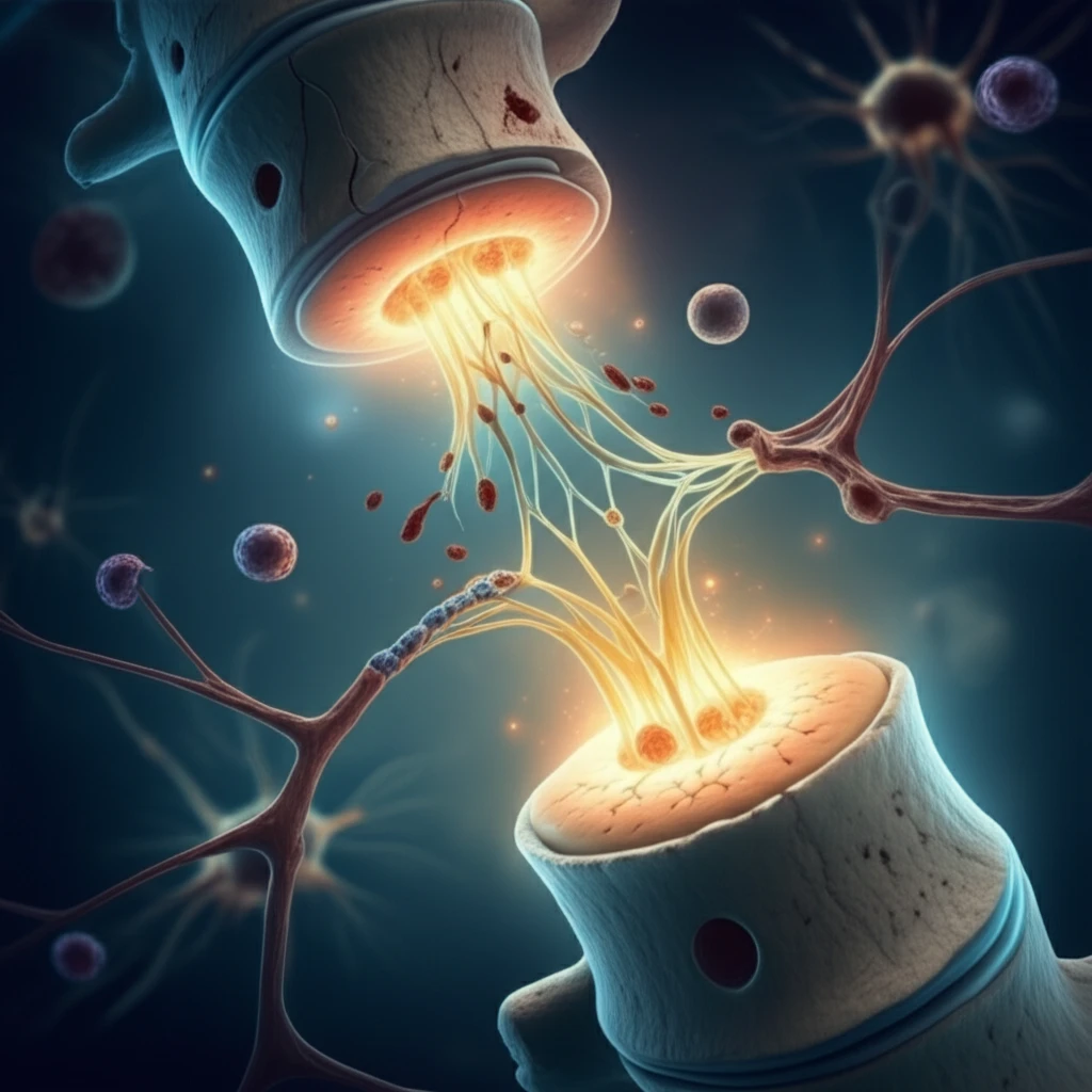
One such promising approach involves the use of peripheral nerve grafts (PNGs). These grafts act as a scaffold, guiding regenerating nerve fibers across the injury site. When combined with acidic fibroblast growth factor (FGF1), a protein known to promote nerve growth and survival, the potential for functional recovery is significantly enhanced. However, successful implementation of this strategy hinges on the precise placement of these PNGs, a challenge that has spurred the development of innovative delivery methods.

Now, scientists are exploring a groundbreaking solution: a biodegradable device that precisely positions PNGs and delivers FGF1 directly to the injury site. This innovative approach aims to overcome previous limitations and offer a more effective and translatable treatment for spinal cord injuries.

The Science Behind the Spinal Cord Repair Breakthrough

Illustration of a biodegradable device repairing a damaged spinal cord, symbolizing nerve regeneration and restored function.

The study, published in Restorative Neurology and Neuroscience, details the development and testing of this novel biodegradable device. Researchers at the Karolinska Institute in Stockholm, Sweden, led by Jonathan Nordblom and Mikael Svensson, designed the device to address the challenges associated with traditional PNG implantation.

The core innovation lies in the device's ability to provide structural support for the PNGs while simultaneously delivering FGF1, a growth factor crucial for nerve regeneration. The device is constructed from calcium sulphate, a biocompatible material that slowly degrades over time, eliminating the need for surgical removal and minimizing long-term complications within the central nervous system (CNS).

  • Peripheral Nerve Grafts (PNGs): Act as a physical bridge, guiding regenerating axons across the damaged spinal cord segment.
  • Acidic Fibroblast Growth Factor (FGF1): A potent growth factor that promotes nerve cell survival, proliferation, and axon extension.
  • Biodegradable Device: Provides structural support for the PNGs, ensures precise placement, and allows for controlled release of FGF1 directly at the injury site.
  • Calcium Sulphate Composition: Guarantees biocompatibility and gradual degradation of the device, minimizing the risk of long-term complications.
The study involved creating a spinal cord resection injury in rats, followed by implantation of the biodegradable device containing PNGs, with or without FGF1. Over several weeks, the animals were assessed using a combination of behavioral testing (BBB score), electrophysiology (motor evoked potentials or MEPs), and immunohistochemistry to evaluate the extent of nerve regeneration and functional recovery.

Looking Ahead: What This Means for Future Spinal Cord Injury Therapies

This research represents a significant step forward in the development of effective therapies for spinal cord injuries. The biodegradable device offers a promising platform for delivering targeted regenerative therapies directly to the injury site. While further research is needed to optimize the device design and FGF1 delivery, these findings pave the way for future clinical trials and ultimately, improved outcomes for individuals living with spinal cord injuries.

About this Article -

This article was crafted using a human-AI hybrid and collaborative approach. AI assisted our team with initial drafting, research insights, identifying key questions, and image generation. Our human editors guided topic selection, defined the angle, structured the content, ensured factual accuracy and relevance, refined the tone, and conducted thorough editing to deliver helpful, high-quality information.See our About page for more information.

Everything You Need To Know

1

What is a peripheral nerve graft (PNG) and why is it used in spinal cord injury treatment?

A peripheral nerve graft (PNG) acts as a scaffold to guide regenerating nerve fibers across the injury site in the spinal cord. It's crucial because the spinal cord's ability to naturally regenerate is limited, and the PNG provides a physical pathway for nerve axons to regrow across the damaged area. The implications of this approach are significant, as it offers a way to overcome the body's natural barriers to spinal cord repair, potentially restoring lost motor and sensory functions.

2

What is acidic Fibroblast Growth Factor (FGF1), and what role does it play in helping to repair spinal cord damage?

Acidic Fibroblast Growth Factor (FGF1) is a protein that promotes nerve cell survival, proliferation, and axon extension. In spinal cord injury treatment, FGF1 is important because it enhances the regenerative capacity of damaged nerves. The presence of FGF1 can significantly improve the effectiveness of peripheral nerve grafts (PNGs), leading to better functional recovery. However, FGF1 alone is not sufficient; it needs to be delivered in a targeted manner, often in conjunction with a structural support system.

3

What is the biodegradable device and how does it improve spinal cord regeneration?

The biodegradable device is designed to provide structural support for peripheral nerve grafts (PNGs) and ensure precise placement at the injury site, while also allowing for controlled release of acidic Fibroblast Growth Factor (FGF1). Its importance lies in overcoming the limitations of traditional PNG implantation techniques. By degrading over time, it minimizes the risk of long-term complications within the central nervous system (CNS). The implications of using a biodegradable device include reduced need for additional surgeries and a more targeted approach to drug delivery.

4

Why is the biodegradable device made of calcium sulphate?

Calcium sulphate is used in the biodegradable device because it is a biocompatible material that slowly degrades over time within the body. This biocompatibility minimizes the risk of adverse reactions or rejection by the body's immune system. The gradual degradation of calcium sulphate eliminates the need for surgical removal of the device, further reducing the potential for complications in the central nervous system (CNS). The implications are significant as it allows for a less invasive and more controlled approach to spinal cord regeneration.

5

Why combine peripheral nerve grafts (PNGs) with acidic Fibroblast Growth Factor (FGF1)?

The combination of peripheral nerve grafts (PNGs) with acidic Fibroblast Growth Factor (FGF1) offers a synergistic approach to spinal cord repair. The PNGs provide a physical scaffold for nerve regeneration, while FGF1 promotes nerve cell survival and growth. This combined approach is more effective than using either method alone. Precise placement of PNGs, facilitated by a biodegradable device, further enhances the regenerative potential. The combined strategy is important to achieve meaningful functional recovery after spinal cord injury.

Newsletter Subscribe

Subscribe to get the latest articles and insights directly in your inbox.