Amplatzer Vascular Plug blocking errant chest tube tract in liver.

When Chest Tubes Go Wrong: How a Vascular Plug Can Save the Day

"A minimally invasive technique offers a solution when a chest tube accidentally enters the liver."


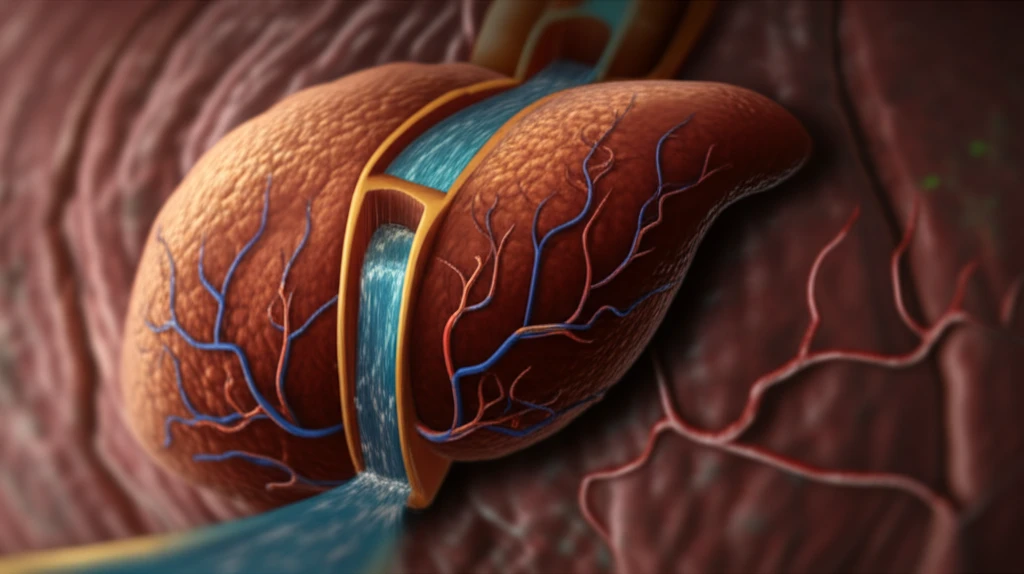
Chest tubes are essential tools in managing various thoracic conditions, such as pleural effusions and pneumothoraxes. However, the insertion of these tubes isn't always straightforward, and complications can arise, including misplacement. While rare, one particularly dangerous complication occurs when a chest tube inadvertently punctures the liver. This can lead to significant bleeding and require immediate intervention to prevent life-threatening consequences.

A recent case report highlighted in Cardiovascular and Interventional Radiology details a scenario where a 70-year-old man experienced this exact complication. Following chest tube insertion to address a collapsed lung and pleural effusion, the tube was found to have penetrated the liver, traveling through the hepatic vein and reaching the right ventricle of the heart. This prompted an emergency response and a clever solution from interventional radiologists.

Traditionally, managing such complications would involve more invasive surgical procedures. However, advancements in interventional radiology have paved the way for less invasive approaches. The article we're diving into showcases the use of an Amplatzer Vascular Plug (AVP) to effectively embolize, or block, the misplaced chest tube tract in the liver. This technique offers a minimally invasive alternative to open surgery, reducing patient risk and recovery time.

The Case of the Misplaced Chest Tube: A Step-by-Step Rescue

Amplatzer Vascular Plug blocking errant chest tube tract in liver.

The initial chest tube insertion in the 70-year-old patient led to a rapid loss of blood, triggering cardiopulmonary resuscitation. Imaging revealed the alarming path of the chest tube through the liver and into the heart. Fortunately, the patient's condition was stabilized, allowing time for a planned intervention.

The interventional radiology team took a meticulous approach:

  • Careful Withdrawal: Under fluoroscopic guidance, the chest tube was carefully withdrawn to the inferior vena cava (IVC), using the patient's vertebral contours as landmarks.
  • Guidewire and Catheter Placement: A 0.035-inch hydrophilic guidewire and a 5-Fr catheter were advanced through the chest tube into the IVC, then pulled back into the hepatic vein, creating a pathway for the AVP.
  • AVP Deployment: A 6-mm Amplatzer vascular plug (AVP) IV was advanced through the catheter and strategically placed to block the liver tract. The AVP was initially positioned in the hepatic vein and then pulled back to the hepatic parenchyma.
  • Confirmation and Release: The team used a nearby liver calcification and patient feedback to estimate the vein-tract junction and ensure correct AVP placement. Once satisfied that there was no bleeding, the AVP was detached, and the chest tube and catheter were removed.
The patient recovered well and went on to receive lung cancer treatment for over a year, highlighting the importance of effectively managing complications to allow for further necessary medical care. The AVP embolization provided a stable foundation for the patient's overall treatment plan.

The AVP Advantage: A Safe and Effective Alternative

The use of AVPs to embolize errant chest tube tracts appears to be a safe and effective method, especially when compared to more invasive surgical options. The minimally invasive nature of the procedure translates to reduced patient risk, shorter recovery times, and a decreased likelihood of complications. However, it's important to note that patients with hepatic congestion may require alternative approaches, such as occlusive balloons, to manage bleeding effectively.

About this Article -

This article was crafted using a human-AI hybrid and collaborative approach. AI assisted our team with initial drafting, research insights, identifying key questions, and image generation. Our human editors guided topic selection, defined the angle, structured the content, ensured factual accuracy and relevance, refined the tone, and conducted thorough editing to deliver helpful, high-quality information.See our About page for more information.

Everything You Need To Know

1

What is an Amplatzer Vascular Plug (AVP), and how is it used in the context of chest tube complications?

An Amplatzer Vascular Plug (AVP) is a device used by interventional radiologists to embolize, or block, blood vessels. In the case of chest tube misplacement where the tube inadvertently punctures the liver, an AVP can be deployed to block the tract created by the chest tube within the liver. This minimally invasive technique prevents further bleeding and avoids the need for more invasive surgical procedures. The AVP effectively seals off the damaged vessel, promoting healing and stability.

2

What are the potential complications of a misplaced chest tube, and why is it important to address them quickly?

A misplaced chest tube, particularly one that punctures the liver, can lead to significant internal bleeding and potentially life-threatening consequences. In a specific case, a chest tube traveled through the hepatic vein and into the right ventricle of the heart. Addressing these complications quickly is crucial to stabilize the patient, prevent further damage, and allow for subsequent medical treatments, such as lung cancer therapy. Rapid intervention with techniques like Amplatzer Vascular Plug (AVP) embolization can minimize risks and improve patient outcomes.

3

How does the use of Amplatzer Vascular Plugs (AVPs) compare to traditional surgical methods for correcting chest tube misplacements in the liver?

Traditional surgical methods for correcting chest tube misplacements in the liver are typically more invasive, carrying higher patient risk, longer recovery times, and an increased likelihood of complications. In contrast, using Amplatzer Vascular Plugs (AVPs) offers a minimally invasive alternative. The AVP procedure involves a small puncture and targeted embolization of the errant chest tube tract, leading to reduced patient trauma, quicker recovery, and a more favorable outcome. However, cases with hepatic congestion might necessitate alternative approaches like occlusive balloons.

4

Can you elaborate on the step-by-step procedure of using an Amplatzer Vascular Plug (AVP) to correct a misplaced chest tube that has entered the liver?

The procedure involves several key steps. First, the chest tube is carefully withdrawn under fluoroscopic guidance to the inferior vena cava (IVC). Next, a guidewire and catheter are advanced through the chest tube into the IVC and then pulled back into the hepatic vein, creating a pathway. An Amplatzer Vascular Plug (AVP) is then advanced through the catheter and strategically placed to block the liver tract, initially positioned in the hepatic vein and then pulled back to the hepatic parenchyma. Finally, using landmarks and patient feedback, the team confirms correct AVP placement and ensures there is no bleeding before detaching the AVP and removing the chest tube and catheter. This meticulous approach ensures effective embolization and minimizes the risk of complications.

5

What factors might make a patient unsuitable for Amplatzer Vascular Plug (AVP) embolization, and what alternative treatments are available?

Patients with hepatic congestion may not be suitable candidates for Amplatzer Vascular Plug (AVP) embolization. In such cases, alternative treatments like occlusive balloons can be used to manage bleeding effectively. Occlusive balloons provide temporary blockage, allowing for clot formation and preventing further hemorrhage. The choice of treatment depends on the specific clinical situation and the patient's overall health status, with interventional radiologists carefully assessing the best approach to minimize risks and maximize the chances of a successful outcome.

Newsletter Subscribe

Subscribe to get the latest articles and insights directly in your inbox.