
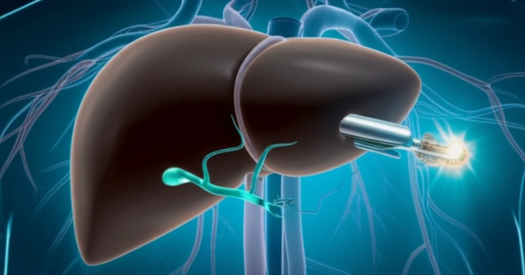
When Chest Tubes Go Wrong: A Simple Plug Can Save the Day

"Learn how a common vascular plug can effectively resolve accidental chest tube placements in the liver, preventing severe complications."
Medical procedures, while intended to heal, occasionally take unforeseen turns. One such complication arises when inserting a chest tube, a common procedure to drain fluid or air from the chest cavity. In rare instances, the tube can be misdirected, entering organs like the liver, leading to significant bleeding and potentially life-threatening situations.
A recent case highlighted by Hamanaka et al. [1] underscores this challenge, prompting physicians to share similar experiences and innovative solutions. One such solution involves the use of Amplatzer Vascular Plugs (AVP), typically used to block blood vessels, to seal off the accidental tract created by the misplaced chest tube.
This approach offers a minimally invasive way to stop the bleeding and stabilize the patient, avoiding more complex surgical interventions. This article explores how AVPs are used in these emergency situations, providing insights into the procedure, its benefits, and considerations for patient care.
Scope of Chest Tube Complications
Chest tubes are among the most commonly placed invasive devices in hospital settings, used to manage pleural effusions, pneumothoraxes, and post-surgical drainage. While generally safe, complications—particularly misplacement—remain a recognized clinical concern. Misplaced chest tubes can cause severe internal organ injury, including penetration of the liver, and may lead to life-threatening hemorrhage requiring urgent intervention. Precise complication rates vary across studies and populations, but the potential for serious harm underscores the need for reliable management strategies.
Chest Tube Misplacement and Embolization Solutions
Chest tubes are essential tools in managing thoracic conditions such as pleural effusions and pneumothoraxes, but their insertion is not always straightforward and complications can arise, including misplacement into adjacent organs. One particularly dangerous scenario occurs when a chest tube is malpositioned into the liver, which can cause severe bleeding and requires immediate intervention. Interventional radiologists have increasingly used Amplatzer Vascular Plugs (AVP) to embolize errant chest tube tracts as a minimally invasive technique that quickly stops bleeding and stabilizes the patient. In one documented case, balloon-assisted hepatic tract embolization using a pull-through technique was successfully performed to remove a malpositioned chest tube that had penetrated the liver and extended into the right ventricle. This approach represents a less invasive alternative to open surgery for managing such complications.
Evolution of Vascular Plugs and Embolization
The advent of vascular plugs for peripheral embolization dates to the early part of this century, marking a significant milestone in interventional medicine. The Amplatzer Vascular Plug (AVP) was created for peripheral embolization as a modification of the family of Amplatzer septal occluders used in the treatment of congenital heart malformations. Over the decades, the development of embolic materials has been key in modern embolization techniques, with major milestones improving how vascular conditions are treated. The family of vascular plugs has expanded over time, with newer variants offering lower profiles and the ability to be delivered through diagnostic catheters. These plugs are ideally suited to close extra-cardiac, high-flow vascular communications and have become versatile and easy to use.
The Case of the Misplaced Chest Tube: A Bleeding Emergency
Consider the case of a 70-year-old man presenting with dyspnea (shortness of breath). Initial diagnostics, including a computed tomography (CT) scan, revealed a mass in the right lung that appeared to be invading the chest wall. The patient also had a collapsed right upper lobe and a pleural effusion (fluid accumulation in the chest cavity).
- The Challenge: Misplaced chest tubes can cause severe bleeding and require immediate intervention.
- The Solution: Interventional radiologists are using Amplatzer Vascular Plugs (AVP) to embolize the errant chest tube tract in the liver.
- The Outcome: This minimally invasive approach quickly stops bleeding and stabilizes the patient.
Current State of Embolization Research
Research into vascular embolization techniques continues to evolve, with recent studies evaluating innovations in plug technology and new device platforms. The broader field of vascular embolization is experiencing sustained growth, driven by increasing use of minimally invasive procedures across multiple clinical specialties. Advances in device design and deployment precision are shaping the direction of ongoing investigations, though comprehensive long-term outcome data remain an area of active development.
Challenges with Micro Vascular Plugs in Peripheral Arterial Embolization
A three-year study examining the use of Micro Vascular Plugs (MVP) in peripheral arterial embolization investigated factors including the type of vascular injury, anticoagulation therapy status at the time of procedure, anatomical district, target artery caliper, landing zone characteristics, and use of additional embolics. The study assessed both technical and clinical success rates, providing real-world data on the performance of these devices. While the MVP system offers a dedicated plug option for smaller vessels, outcomes can be influenced by patient-specific factors such as anticoagulation status and vessel anatomy. These findings highlight that device selection and patient characteristics play important roles in procedural success, and not all cases achieve optimal results uniformly.
Tract Embolization and Chest Tube Management Practices
Research comparing tract embolization versus no embolization after CT-guided percutaneous lung biopsy has evaluated whether tract embolization is effective in preventing pneumothorax and subsequent chest tube placement. Comprehensive reviews of chest tube management have assessed existing research to highlight best practices and safe application, providing direction for future clinical use. In the context of variceal embolization, studies have directly compared vascular plugs and pushable coils, examining the effectiveness of each device type in achieving complete occlusion of blood flow. The operator's choice between plug types and additional sclerosant use can vary, suggesting that procedural decision-making remains an important variable across embolization applications.
AVP: A Safe and Effective Solution
In summary, the use of Amplatzer Vascular Plugs appears to be a safe and effective method for embolizing errant tracts created by malpositioned chest tubes. While this technique offers a promising solution, it's crucial to consider individual patient factors. Patients with hepatic congestion, for instance, might benefit more from an occlusive balloon technique. Further research and clinical experience will continue to refine and optimize the use of AVPs in these challenging situations, ultimately improving patient safety and outcomes.
Vascular Plug Embolization: Outcomes and Device Evolution
Case reports have documented instances of vascular plug embolization requiring computed tomography angiography of the abdominal aorta and iliofemoral system with bilateral leg runoffs for evaluation, demonstrating that device migration remains a recognized complication. The evolution of the Amplatzer Vascular Plug family has been extensively reviewed, with attention to current applications and device refinements. Clinicians using these devices must weigh the benefits of minimally invasive tract embolization against the risk of device-related complications, integrating case-level evidence into broader practice decisions.
Market Growth and Technological Trajectory in Vascular Embolization
The vascular embolization market is projected to grow from approximately USD 2.09 billion in 2024 to USD 5.9 billion by 2035, at a compound annual growth rate of 9.9%. Major trends in the forecast period include increasing use of minimally invasive embolization procedures, rising adoption of removable and adjustable vascular plugs, and growing demand for precise vessel occlusion devices. Expansion of catheter-based interventions and enhanced focus on improved deployment control are also expected to shape the market. These projections suggest sustained investment and innovation in the vascular plug and embolization device space over the coming decade.
Systemic Considerations in Chest Tube Complication Management
The management of chest tube complications sits at the intersection of procedural skill, device technology, and systemic healthcare capacity. Access to interventional radiology expertise and appropriate embolization equipment varies significantly across healthcare settings, creating disparities in patient outcomes. Training standardization and protocol development remain ongoing challenges as minimally invasive techniques expand in scope and adoption. Addressing these systemic factors will be important to ensuring that innovations like vascular plug tract embolization benefit patients broadly rather than only in well-resourced centers.
Patient Safety and Clinical Decision-Making
When a chest tube is misplaced, the clinical team must act rapidly to assess the extent of organ injury and choose the most appropriate intervention. The shift toward minimally invasive embolization techniques reflects a broader movement in medicine to reduce procedural morbidity while achieving effective treatment. Patient outcomes in these scenarios depend not only on device performance but also on timely recognition of complications and the availability of trained specialists. The human factors of clinical judgment, communication, and team coordination remain central to managing these high-stakes situations safely.