Brain bathed in sunlight surrounded by calcium symbols and mitochondria.

Vitamin D: The Brain Booster You Didn't Know You Needed?

"New research suggests vitamin D could play a key role in brain repair and calcium regulation, offering hope for multiple sclerosis treatment."


Low vitamin D levels have long been recognized as an environmental risk factor for multiple sclerosis (MS). Supplementation with vitamin D appears to reduce disease activity in MS patients. Vitamin D signaling, mainly mediated through the vitamin D receptor (VDR), influences various cells in the central nervous system (CNS), including oligodendrocytes, astrocytes, microglia, and immune cells. This has spurred interest in understanding exactly how vitamin D impacts brain health, especially in demyelinating diseases like MS.

Scientists have been investigating the potential of vitamin D to promote remyelination—the repair of damaged myelin sheaths that protect nerve fibers—using the cuprizone model, an animal model that mimics demyelination and spontaneous remyelination. Prior research indicates that 1,25-dihydroxyvitamin-D3 (1,25D), the hormonally active form of vitamin D, may stimulate remyelination and oligodendrocyte maturation, which are key to nerve repair.

A new study dives deeper, aiming to identify how 1,25D treatment affects the brain's overall protein landscape during remyelination. By understanding which proteins are influenced by 1,25D, researchers hope to uncover signaling mechanisms that could be harnessed to improve remyelination in MS patients. This is particularly relevant as several clinical trials are underway to assess the impact of vitamin D intervention in MS, making the identification of key proteins crucial for monitoring treatment effects.

Decoding Vitamin D's Impact on Brain Proteins: What the Study Revealed

Brain bathed in sunlight surrounded by calcium symbols and mitochondria.

Researchers employed a technique called quantitative proteomics to analyze brain tissue from mice treated with cuprizone and supplemented with high-dose 1,25D or a placebo. This method allowed them to quantify thousands of proteins and identify those that were differentially regulated by 1,25D during remyelination. The scientists quantified 5062 proteins, pinpointing 125 that exhibited significant changes in expression due to the 1,25D treatment.

The study revealed compelling patterns in protein regulation:

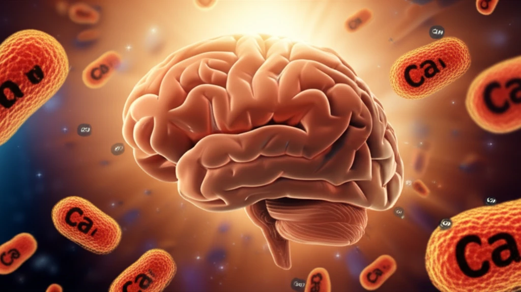
  • Early Remyelination Boost: Proteins involved in calcium binding, such as calretinin, S10A5, and secretagogin, were upregulated in the early phases of remyelination. These proteins play crucial roles in calcium homeostasis and signaling within cells.
  • Mitochondrial Support: Proteins linked to mitochondrial function, including NADH-ubiquinone oxidoreductase chain 3 and acyl-coenzyme A synthetase, also showed increased activity during the early remyelination phase. Mitochondria are essential for energy production and overall cell health.
  • Calretinin Spotlight: Further analysis using immunohistochemistry confirmed that calretinin immunoreactivity was significantly increased in the medial septal nuclei of mice treated with 1,25D during early remyelination, highlighting its importance in the observed effects.
These findings suggest that vitamin D may influence remyelination through mechanisms involving increased expression of calretinin and potentially other calcium-binding proteins. This indicates a targeted approach where vitamin D supplementation can modulate specific proteins to enhance brain repair.

What Does This Mean for MS and Brain Health?

This research provides valuable insights into how vitamin D may impact brain repair processes, particularly in the context of demyelinating diseases like multiple sclerosis. By identifying specific proteins regulated by 1,25D, the study points towards potential therapeutic targets for enhancing remyelination.

The upregulation of calcium-binding proteins like calretinin suggests that vitamin D could play a critical role in maintaining calcium homeostasis and promoting cell signaling pathways that support nerve repair. Similarly, the influence on mitochondrial proteins highlights the importance of energy production and cellular health in the remyelination process.

While further research is needed to fully understand the implications of these findings, this study offers a promising foundation for developing vitamin D-based strategies to improve brain health and potentially alleviate the symptoms of MS. Keeping vitamin D levels in check through diet, supplementation, and sunlight exposure might be a proactive step in supporting overall neurological well-being.

About this Article -

This article was crafted using a human-AI hybrid and collaborative approach. AI assisted our team with initial drafting, research insights, identifying key questions, and image generation. Our human editors guided topic selection, defined the angle, structured the content, ensured factual accuracy and relevance, refined the tone, and conducted thorough editing to deliver helpful, high-quality information.See our About page for more information.

This article is based on research published under:

DOI-LINK: 10.1016/j.neuint.2017.08.008, Alternate LINK

Title: 1,25-Dihydroxyvitamin-D3 Induces Brain Proteomic Changes In Cuprizone Mice During Remyelination Involving Calcium Proteins

Subject: Cell Biology

Journal: Neurochemistry International

Publisher: Elsevier BV

Authors: Eystein Oveland, Agnes Nystad, Frode Berven, Kjell-Morten Myhr, Øivind Torkildsen, Stig Wergeland

Published: 2018-01-01

Everything You Need To Know

1

How does Vitamin D impact multiple sclerosis (MS) and the central nervous system (CNS)?

Vitamin D, particularly its hormonally active form 1,25-dihydroxyvitamin-D3 (1,25D), appears to reduce disease activity in multiple sclerosis (MS) patients. It influences cells in the central nervous system (CNS), like oligodendrocytes, astrocytes, microglia, and immune cells. Scientists are exploring its potential to promote remyelination, the repair of damaged myelin sheaths, using the cuprizone model. 1,25D may stimulate remyelination and oligodendrocyte maturation, which are essential for nerve repair.

2

What were the key methods and protein findings of the recent study on Vitamin D and brain health?

The study employed quantitative proteomics to analyze brain tissue from mice treated with cuprizone and supplemented with high-dose 1,25D or a placebo. They quantified 5062 proteins and found that 125 exhibited significant changes in expression due to the 1,25D treatment. Proteins involved in calcium binding, such as calretinin, S10A5, and secretagogin, were upregulated early in remyelination. Additionally, proteins linked to mitochondrial function, including NADH-ubiquinone oxidoreductase chain 3 and acyl-coenzyme A synthetase, showed increased activity. Immunohistochemistry confirmed increased calretinin immunoreactivity in the medial septal nuclei of mice treated with 1,25D.

3

What implications does this research have for treating multiple sclerosis and promoting overall brain health?

The research highlights the potential of vitamin D to impact brain repair processes, especially in demyelinating diseases like multiple sclerosis. By identifying specific proteins regulated by 1,25D, the study suggests potential therapeutic targets for enhancing remyelination. Specifically, it points towards mechanisms involving increased expression of calretinin and other calcium-binding proteins, indicating a targeted approach where vitamin D supplementation can modulate specific proteins to enhance brain repair. These findings are particularly relevant as several clinical trials are underway to assess the impact of vitamin D intervention in MS, making the identification of key proteins crucial for monitoring treatment effects.

4

What role do calcium-binding proteins like calretinin play in the remyelination process?

Calretinin, S10A5, and secretagogin are calcium-binding proteins that were found to be upregulated during the early phases of remyelination in the study. These proteins are essential for calcium homeostasis and signaling within cells. The increased expression of calretinin, in particular, was confirmed through immunohistochemistry in the medial septal nuclei of mice treated with 1,25D. These proteins play a critical role in the observed effects of Vitamin D and point toward the ability of Vitamin D to modulate specific proteins to enhance brain repair.

5

How does Vitamin D affect mitochondrial function and energy production in the context of brain repair?

Mitochondrial function is supported by proteins like NADH-ubiquinone oxidoreductase chain 3 and acyl-coenzyme A synthetase, which showed increased activity during the early remyelination phase. Mitochondria are essential for energy production and overall cell health. By upregulating these proteins, vitamin D supplementation may enhance the energy supply and metabolic functions necessary for effective remyelination. This aspect connects to the larger context of the study, indicating that the beneficial effects of Vitamin D involve not just direct nerve repair, but also supporting cellular functions that enable that repair.

Newsletter Subscribe

Subscribe to get the latest articles and insights directly in your inbox.