
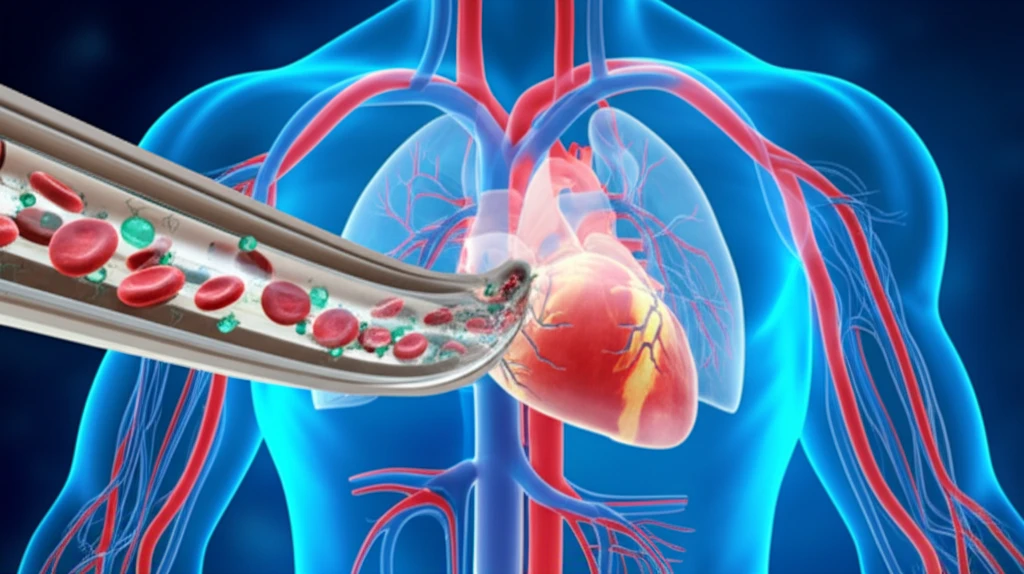
Illustration of a microfluidic device, vein-on-a-chip, with cells, highlighting its role in cardiovascular research and drug discovery.

Vein-on-a-Chip: A Revolutionary Approach to Cardiovascular Research

"Scientists Develop Microfluidic Device Mimicking Blood Vessels for Advanced Cell Culture and Drug Discovery"


Cardiovascular diseases remain a leading cause of death worldwide, underscoring the critical need for advanced research methods. Traditional cell culture techniques and animal models often fall short in accurately replicating the complex environment of human blood vessels. Now, a team of scientists has developed an innovative "vein-on-a-chip" microfluidic device, offering a promising new approach to cardiovascular research.

This "vein-on-a-chip" technology mimics the structure and function of a blood vessel, allowing for cell culture under controlled conditions. By using polyester-toner microchips, the researchers created a platform that enables a deeper understanding of vascular health, drug effects, and the mechanisms behind diseases like atherosclerosis. This advancement holds tremendous potential for revolutionizing drug discovery and personalized medicine.

The simplicity and effectiveness of this new device highlights its potential to accelerate the pace of discoveries in cell biology and organ-on-a-chip research. This technology is not just a scientific achievement, but also a testament to the power of innovation in addressing pressing global health challenges.

AI Search Multiple angles on this topic

Current Research Impact

Marder et al. engineered an open microfluidic vessel-on-chip platform for multiomics cell analysis, showing that flow induction supported the formation and maturation of functional vessels from stem cell-derived endothelial cells. Applying oxidative stress to these vessels-on-chip enabled investigation of the early onset of cardiovascular disease. Separately, scientists at the University of Birmingham developed a vein-on-a-chip model to study mechanisms of blood-clot formation, with potential to reduce reliance on animals in cardiac-disease research.

Limits of Conventional Models

Traditional in vitro models remain widely used to study vascular diseases such as atherosclerosis and thrombosis, but the cited reviews report that they do not fully recapitulate the complexity of the in vivo microenvironment. Vessel-on-a-chip research therefore examines microvascular transport and physiology while evaluating different microfabrication strategies. These systems are presented as a promising alternative because they can incorporate multiple cell types and more closely model vascular conditions, although current challenges and limitations remain.

Building the Vein-Chip

A foundational development was the creation of a microengineered human Vein-Chip using organ-on-a-chip methodology. The platform integrated fully vascularized microfluidic channels containing venous-valve architecture and enabled whole-human-blood perfusion. This work established a model designed to reproduce important structural and flow-related features of human veins for vascular research.

How the "Vein-on-a-Chip" Works: A Detailed Look at the Technology

Illustration of a microfluidic device, vein-on-a-chip, with cells, highlighting its role in cardiovascular research and drug discovery.

The "vein-on-a-chip" is a microfluidic device designed to mimic the environment inside a blood vessel. The device uses a polyester-toner microchip, which includes a channel that serves as a miniaturized blood vessel. This channel is about 2 centimeters long and 0.2 centimeters wide, designed to allow for cell culture under conditions that closely replicate the physiological environment within the human body.

The fabrication process is relatively straightforward. The microchips are created using a print, cut, and laminate (PCL) approach, which involves printing a channel design onto polyester films using a laser printer. The channel depth is controlled by the thickness of the toner layer. The chip is then treated with oxygen plasma and fibronectin, promoting cell adhesion and growth. Researchers use HUVEC cells (Human Umbilical Vein Endothelial Cells) and these are cultured within the microchannel, providing an environment suitable for cell studies, drug testing, and toxicological research.

  • Oxygen Plasma Treatment: Enhances surface properties for better cell adhesion.
  • Fibronectin Coating: Provides a matrix for cells to attach and grow.
  • Continuous Perfusion: Maintains a dynamic environment, mimicking blood flow.
  • Endpoint Assays: Methods to evaluate cell viability and nitric oxide production.
AI Search Multiple angles on this topic

Emerging Evidence

Recent work suggests that vein-on-a-chip and vessel-on-a-chip platforms are becoming useful tools for studying vascular biology and cardiovascular disease. However, the available material indicates an emerging research area rather than a settled clinical technology. Further studies will be needed to determine how consistently these models reproduce human disease and support practical research applications.

Remaining Uncertainties

The promise of vein-on-a-chip technology does not eliminate the challenges associated with reproducing the full complexity of human vascular biology. Models may capture selected features of blood flow, vessel structure, or disease processes without representing every relevant biological interaction. Their findings should therefore be interpreted as complementary experimental evidence rather than automatic substitutes for all established methods.

Model Comparison

Vein-on-a-chip systems offer a potentially more physiologically relevant way to study vascular processes than simplified conventional culture models. Their main comparative value lies in combining controlled microfluidic flow with vascular structures and, in some designs, human blood or multiple cell types. The extent of their advantage will depend on how faithfully each platform reproduces the specific disease mechanism under investigation.

The key to the device's effectiveness is its ability to control various factors such as fluid flow and pressure, which create a microenvironment that closely mimics physiological conditions. This allows researchers to study how cells respond to different stimuli and to test the effects of various substances, offering a more accurate and relevant platform for drug discovery and research.

The Future of Cardiovascular Research: Implications and Potential

The "vein-on-a-chip" represents a significant leap forward in cardiovascular research. The ability to create an in-vitro model that accurately reflects the complexities of the human vascular system promises to accelerate the development of new therapies for heart disease and related conditions. The simplicity and cost-effectiveness of the device make it an attractive option for many laboratories, potentially democratizing access to advanced research tools and accelerating the pace of discovery. As the research continues, we can expect to see this technology play a pivotal role in the future of cardiovascular health and personalized medicine.

AI Search Multiple angles on this topic

A Complementary Platform

Taken together, the supplied material portrays vein-on-a-chip technology as a promising research platform rather than a complete replacement for existing cardiovascular methods. Its value comes from combining microfluidic control with vessel-like architecture, cell-based analysis, and disease-relevant stresses such as oxidative conditions or blood perfusion. Expert assessment will ultimately depend on reproducibility, biological fidelity, and demonstrated usefulness in cardiovascular research.

Toward Physiological Models

Recent reviews describe vascular organoids and vessel-on-chip technologies as powerful tools for studying human diseases, including cardiovascular diseases. Vascular organoids generated from human pluripotent stem cells can self-organize into complex three-dimensional structures that more closely mimic human biology. Future research is therefore likely to explore increasingly physiologically relevant combinations of vascular organoids and vessel-on-chip platforms.

Translational Challenges

The broader adoption of vein-on-a-chip research may depend on addressing differences between engineered models and human vascular systems. Standardization, reproducibility, and validation across laboratories are likely to remain important challenges. Until those issues are resolved, promising chip-based findings may be difficult to compare directly or translate into routine medical practice.

Potential Human Benefit

Vein-on-a-chip research could ultimately support better understanding of blood-clot formation and vascular disease. By enabling controlled studies of human-relevant vessel conditions, these models may contribute to improved drug testing and therapeutic discovery. Their real-world impact remains prospective, because the supplied material does not establish clinical outcomes or widespread medical adoption.

How the polyester-toner chip compares with related work

A later review likewise cites oxygen plasma followed by fibronectin as a way to support endothelial-cell adhesion and proliferation on polyester-based vein chips.[1]

The study at a glance, in the paper's own terms

Study designIn vitro (cell or tissue) study[2]
PopulationHUVEC cells cultured in polyester-toner microchips[2]
DurationCultures were viable for 5 consecutive days[2]
Effect sizeShear stress was 4.48 dyn cm−2[2]
Times cited30[2]

What an in-vitro endothelial-cell study can establish

In vitro (cell or tissue) study (level 6 of 8)

Because this was an in-vitro study using cultured HUVECs in polyester-toner microchips, it can show how those cells behaved in the device, not establish outcomes in living people.[2]

The study reports a specific shear-stress condition, but the cultured-cell design alone does not show that the same response would occur across other vascular settings.[2]

What the cell-adhesion results add up to

The surface preparation mattered: treated chips supported more cell adherence and confluence than untreated chips, while untreated cells remained spherical and did not form a confluent layer.[3]

Fibronectin was used to mimic extracellular matrix and encourage cells to adhere and take on an elongated morphology, supporting endothelialization of the device.[3]

The endpoint assays found no nitric oxide in the supernatant after cells were cultured with individual chip components; this is a finding about that assay condition, not evidence of nitric oxide production by the assembled, perfused device.[3]

What keeps the chip results from being settled

  • Oxygen plasma alone produced only brief attachment before cells released from the surface, in both static and dynamic setups.[3]
  • Some treated, perfused chips still lacked complete confluence or uniformly elongated cells after the reported experiment period.[3]
  • After five days, cell detachment was associated with crowding as new cells had limited room to attach.[3]

What the perfusion experiments leave unresolved

  • How consistently do cells align with flow under the chip’s low-shear condition, given that the authors observed elongation but no defined flow-direction alignment?[3]
  • Would different flow conditions change cell orientation, as a cited comparison found different alignment behavior at low and high shear?[3]

Terms behind the perfusion findings

Endothelialization
The paper describes this as mimicking extracellular matrix to improve adhesion and encourage cells to assume an elongated morphology on the device.[3]

About this Article -

Written with AI assistance from published research, and reviewed by the Mystum team. See our About page for more information.

This article is based on research published under:

DOI-LINK: 10.1038/s41598-017-11043-0, Alternate LINK

Title: Endothelial Cell Culture Under Perfusion On A Polyester-Toner Microfluidic Device

Subject: Multidisciplinary

Journal: Scientific Reports

Publisher: Springer Science and Business Media LLC

Authors: Ana Carolina Urbaczek, Paulo Augusto Gomes Carneiro Leão, Fayene Zeferino Ribeiro De Souza, Ana Afonso, Juliana Vieira Alberice, Luciana Teresa Dias Cappelini, Iracilda Zeppone Carlos, Emanuel Carrilho

Published: 2017-09-05

Everything You Need To Know

1

What is a "vein-on-a-chip" and why is it important for cardiovascular research?

The "vein-on-a-chip" is a microfluidic device designed to mimic the structure and function of a blood vessel. It's important because traditional cell culture techniques and animal models often fail to accurately replicate the complex environment of human blood vessels. This device offers a more realistic in-vitro model for studying vascular health, drug effects, and disease mechanisms, particularly for conditions like atherosclerosis. It uses polyester-toner microchips to create a miniaturized blood vessel environment, enabling deeper insights into cardiovascular processes. It doesn't currently replace animal studies, but offers a complementary alternative.

2

How does the "vein-on-a-chip" actually work and what materials are used in its construction?

The "vein-on-a-chip" works by creating a miniaturized blood vessel environment within a microfluidic device. It utilizes a polyester-toner microchip with a channel that mimics a blood vessel. The fabrication involves a print, cut, and laminate (PCL) approach, where a channel design is printed onto polyester films using a laser printer. The chip is then treated with oxygen plasma and coated with fibronectin to promote cell adhesion and growth. HUVEC cells (Human Umbilical Vein Endothelial Cells) are cultured within this microchannel, allowing researchers to control factors like fluid flow and pressure, thus replicating physiological conditions for cell studies and drug testing. This controlled environment allows for Endpoint Assays, to evaluate cell viability and nitric oxide production.

3

What are the key benefits of using a "vein-on-a-chip" compared to traditional research methods?

The "vein-on-a-chip" offers several key benefits. It provides a more accurate in-vitro model of the human vascular system compared to traditional cell culture techniques and animal models. The device's controlled environment, maintained by continuous perfusion, allows researchers to study cell responses to different stimuli and test drug effects in a physiologically relevant manner. Its simplicity and cost-effectiveness make it accessible to more laboratories, potentially accelerating the pace of discovery in cardiovascular research and personalized medicine. It allows a focus on the complex interplay of HUVEC cells.

4

How does oxygen plasma treatment and fibronectin coating contribute to the functionality of the "vein-on-a-chip"?

Oxygen plasma treatment enhances the surface properties of the polyester-toner microchip, leading to better cell adhesion. Fibronectin coating then provides a matrix for cells, specifically HUVEC cells, to attach and grow effectively within the microchannel. Without these treatments, cells would not adhere properly to the chip, compromising the cell culture and hindering accurate study of vascular functions and responses to stimuli.

5

What are the potential future applications of the "vein-on-a-chip" in personalized medicine and drug discovery?

The "vein-on-a-chip" holds significant potential for personalized medicine and drug discovery. Its ability to mimic the human vascular system allows for testing patient-specific cells to predict individual responses to drugs, optimizing treatment strategies. In drug discovery, the device can accelerate the identification of promising therapeutic candidates by providing a more accurate and relevant platform for evaluating drug efficacy and toxicity. This technology may also offer insights into patient specific atherosclerosis treatments. Future uses may include disease modeling for rare vascular diseases.

Newsletter Subscribe

Subscribe to get the latest articles and insights directly in your inbox.