
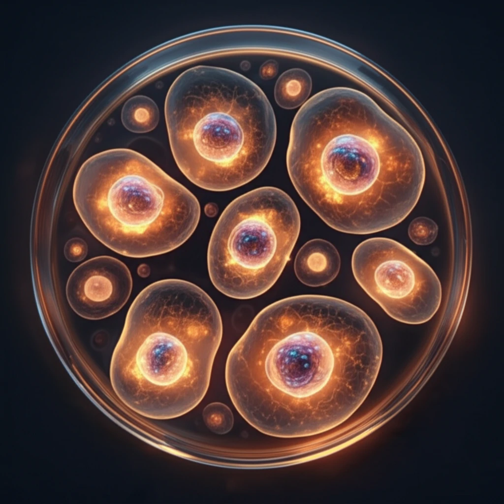
Unlock Bovine Hepatocytes: Revolutionizing Liver Research and Cell Culture

"Discover the secrets to isolating and identifying bovine primary hepatocytes for advanced in vitro studies, enhancing drug metabolism research and toxicity assessments."
The liver, a vital organ with diverse functions, relies heavily on hepatocytes for most of its critical processes. These specialized cells control everything from metabolism to detoxification, making them essential for maintaining overall health. Understanding how hepatocytes function is crucial, and one of the best ways to study them is through in vitro models using primary hepatocytes.
Primary hepatocytes are freshly isolated liver cells that retain many of their original functions, providing a more accurate representation of in vivo conditions than other cell types. They're widely used to study biological processes, evaluate drug toxicity, and investigate metabolic pathways. However, isolating high-quality hepatocytes can be challenging, with traditional methods often resulting in lower cell activity and purity.
Recent research has focused on optimizing hepatocyte isolation techniques to improve cell yield and viability. One promising approach is the two-step collagenase perfusion technique, which involves carefully perfusing the liver with collagenase to break down the extracellular matrix while preserving cell integrity. By identifying key hepatocyte proteins and refining the isolation process, scientists can obtain highly purified and functional cells for a wide range of studies.
Current Hepatocyte Research
Hepatocytes differentiated from induced pluripotent stem cells or stem cells have potential as representative in vitro models of the human liver for research and early safety assessment. Primary human hepatocytes can also be isolated from explanted livers obtained during orthotopic liver transplantation for decompensated cirrhosis or metabolic liver disease without end-stage liver disease. Recent work notes that conventional two-step perfusion isolation requires large samples, substantial resources, specialized expertise, and suitable cannulation vessels, while non-perfusion methods may produce fewer cells and inadequately assess purity.
From Isolation to Culture
A 2025 review describes three decades of human hepatocyte isolation and cryopreservation, emphasizing that human hepatocytes remain an irreplaceable source of functional cells for toxicology and drug development. A 2011 PLOS One study addressed the possibility of isolating hepatocytes from diseased liver tissue removed during transplantation, noting that this could increase cell availability for research. Technical reviews have also documented major milestones in isolating and culturing primary hepatocytes and non-parenchymal liver cell populations, while identifying prolonged in vitro maintenance and proliferation of viable, functional primary hepatocytes as a major challenge.
Optimizing Hepatocyte Isolation: Key Steps and Techniques
The isolation of primary hepatocytes is a delicate process that requires careful attention to detail. The two-step collagenase perfusion technique is a favored method due to its effectiveness in dissociating liver cells while maintaining their functionality. This technique involves two primary steps: a calcium-free perfusion to disrupt cell junctions, followed by collagenase perfusion to digest the connective tissue.
- Initial Perfusion with Calcium-Free Medium: The liver is first perfused with a calcium-free medium, often supplemented with a calcium chelator like EDTA. This step is crucial for disrupting calcium-dependent cell junctions, such as desmosomes, which hold liver cells together.
- Collagenase Perfusion: After the initial perfusion, a collagenase-containing buffer is introduced to digest the extracellular matrix (ECM). The presence of calcium ions is essential during this phase to ensure optimal collagenase activity.
- Mechanical Dispersion: Once the liver tissue has been perfused, it is gently dispersed mechanically, often using a spatula, to further separate the cells. This step helps to break down any remaining connections between cells.
- Filtration and Centrifugation: The resulting cell suspension is filtered to remove connective tissue and debris. Subsequent centrifugation steps separate the hepatocytes from dead cells and non-parenchymal cells, ensuring a purified sample.
Recent Methods and Findings
Recent primary hepatocyte research has examined enzymatic dissociation and pancreatin digestion in marine fish cells cultured in L-15 medium at 17 ± 1 °C for up to six days. Researchers monitored viability and function using trypan blue exclusion, MTT, lactate dehydrogenase activity, and ethoxyresorufin O-deethylase activity after benzo[a]pyrene exposure. In bovine research, isolated calf hepatocytes were identified by confocal immunofluorescence detection of cytokeratin-18, while a rat study reported isolated hepatocyte viability above 95% and purity above 93.2%.
Comparative Experimental Evidence
One comparative liver study examined carbon tetrachloride-induced liver necrosis when hepatocyte proliferation was inhibited by 2-acetylaminofluorene. Morphometric analysis reported equal and maximum liver cell necrosis by 24 hours in experimental and control groups, similar kinetics for removing dead liver cells, and similar mean liver cell area. A separate decellularization study compared porcine and human aortic valves, reporting reduced U-937 migration after decellularization from 832.7×10(3) to 152.4∓31.1×10(3) in porcine valves and from 644.9×10(3) to 91.2×10(3) in human valves, with P<0.01 for both comparisons.
Advancing Hepatocyte Research for Future Applications
The isolation and culture of primary hepatocytes represent a cornerstone in understanding liver function and developing treatments for liver diseases. By optimizing techniques such as the two-step collagenase perfusion method, researchers can obtain high-quality cells for in vitro studies. These studies contribute significantly to drug development, toxicity testing, and our fundamental understanding of liver biology, paving the way for more effective therapies and preventive strategies.
Patient-Derived Hepatocytes
Primary hepatocytes can be successfully isolated from explanted liver specimens obtained from patients receiving orthotopic liver transplantation for decompensated liver cirrhosis. They can also be obtained from patients with metabolic liver disease without end-stage liver disease. These patient-derived cells are described as a valuable resource for pharmaceutical industry research and drug screening.