Unusual Liver Masses? How an Elevated AFP Level Can Signal More Than Just Liver Cancer
"Navigating the complexities of tumor diagnosis when a routine blood test throws you a curveball. Learn how to interpret elevated alpha-fetoprotein levels in the presence of liver masses and the critical steps to take next."
Imagine facing a health scare that begins with vague symptoms like malaise and unexplained weight loss. Routine blood work reveals an elevated alpha-fetoprotein (AFP) level, a marker often associated with liver cancer. Now, picture the added complexity of discovering masses in your liver. The immediate assumption? Hepatocellular carcinoma (HCC), a primary liver cancer. But what if the real culprit is something far less common and initially overlooked?
This is the diagnostic dilemma faced by a 55-year-old man in a recent JAMA Clinical Challenge. His case highlights a crucial lesson for both medical professionals and patients: elevated AFP levels and liver masses don't always equate to HCC. Sometimes, a thorough investigation is needed to uncover the true source of the problem.
In this article, we'll delve into the details of this intriguing case, exploring the diagnostic process, the importance of advanced imaging techniques, and the critical role of biopsy in uncovering the unexpected diagnosis of a pancreatic neuroendocrine tumor (PNET). We'll also discuss how this case underscores the need for vigilance and a broad differential diagnosis when faced with seemingly straightforward medical presentations.
Decoding the Diagnostic Puzzle: When AFP Isn't Just Liver Cancer
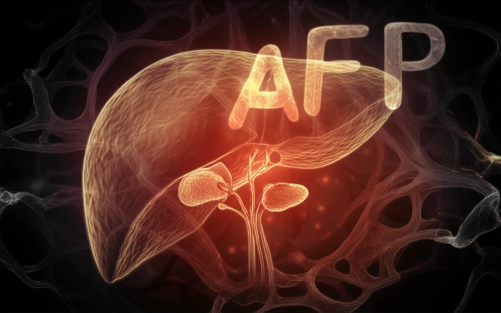
Alpha-fetoprotein (AFP) is a protein produced by the liver and yolk sac during fetal development. Its levels typically drop significantly after birth, with only trace amounts detectable in healthy adults. However, elevated AFP levels can be a sign of certain medical conditions, most notably hepatocellular carcinoma (HCC), the most common type of liver cancer. Because of this strong association, doctors often rely on AFP levels as a key diagnostic marker for HCC, especially in patients with chronic liver disease.
- Comprehensive Medical History and Physical Exam: A thorough assessment helps identify potential risk factors for liver disease (alcohol use, hepatitis history) and other relevant medical conditions.
- Advanced Imaging: Techniques like triple-phase CT scans and MRI can provide detailed information about the characteristics of liver masses, helping to distinguish between different types of tumors.
- Biopsy: This is the gold standard for diagnosis. A tissue sample is taken from the liver mass and examined under a microscope to determine the type of cells present and confirm the diagnosis.
The Takeaway: Think Beyond the Obvious
This case underscores the importance of considering a broad differential diagnosis when evaluating patients with elevated AFP levels and liver masses. While HCC is a common culprit, it's crucial to remember that other conditions can mimic its presentation. Advanced imaging techniques, combined with biopsy, are essential for accurate diagnosis and appropriate treatment. For patients, this means being proactive in discussing all possible diagnoses with your doctor and understanding the rationale behind each diagnostic step. Don't hesitate to ask questions and advocate for thorough investigation, especially when faced with complex medical presentations.