Unraveling the 'Feather Pattern': What It Reveals About Your Muscles
"Discover how this unique muscle imaging sign can be a crucial indicator of underlying muscle diseases and conditions."
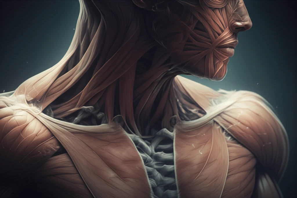
Imagine looking at an image of your muscles and seeing a pattern resembling a feather. This isn't a trick of the light but a real phenomenon known as the 'feather pattern' in muscle imaging. While it might sound unusual, this pattern can be a significant indicator of various muscle diseases and conditions.
The term 'pinnate,' derived from the Latin 'pinnatus,' meaning feathered, describes muscles with fascicles attaching obliquely to a tendon, naturally resembling a feather. However, the 'feather pattern' we're discussing here arises when this arrangement becomes accentuated due to specific pathological changes within the muscle.
In this article, we will explore what the 'feather pattern' signifies, how it is identified through different imaging techniques, and what conditions can cause it. Understanding this sign can lead to earlier and more accurate diagnoses, ultimately improving patient care.
Decoding the Feather Pattern: What Does It Mean?
The 'feather pattern' becomes prominent when the muscle's usual structure is altered, causing the fascicles (muscle fibers) to separate. This separation can be due to several reasons, each detectable through different imaging modalities.
- X-rays: In cases of subcutaneous emphysema, where gas dissects between muscle fascicles, an X-ray can reveal the feather pattern. This typically occurs when air gets trapped under the skin, separating the muscle fibers.
- MRI (Magnetic Resonance Imaging): MRI is particularly effective in visualizing the feather pattern. Muscle atrophy, or the wasting away of muscle tissue, leads to an increase in fat between the fascicles. On T1-weighted MR images, this appears as a distinct feather pattern. Additionally, in grade 1 muscle tears, oedema (swelling) accentuates the pattern on water-weighted/T2 sequences. However, more severe muscle tears will disrupt this pattern.
- Ultrasound: Even with early ultrasound machines, the muscle fascicles can be depicted, revealing a feather-like appearance. This makes ultrasound a readily accessible tool for initial assessments.
The Broader Implications of the Feather Pattern
The identification of the 'feather pattern' is more than just an interesting observation; it's a crucial diagnostic clue. Recognizing this pattern enables healthcare professionals to identify early-stage muscle damage, differentiate between various muscle conditions, and tailor treatment plans more effectively. As imaging technology advances, our ability to detect and interpret these subtle signs will undoubtedly improve, leading to better outcomes for patients with muscle disorders. Staying informed about such diagnostic markers empowers individuals to engage more proactively in their healthcare, leading to earlier detection and management of potential issues.