Unlocking the Secrets Within: How Cellular GPS Influences Health and Disease
"Delving into the hidden world of subcellular localization of the (Pro)renin receptor ((P)RR/ATP6ap2), revealing its profound impact beyond blood pressure regulation."
For years, the renin-angiotensin system (RAS) was understood as a straightforward system for controlling blood pressure through circulating hormones. Renin, an enzyme within this system, was thought to be the active player, while its precursor, prorenin, was considered inactive. This view, however, has dramatically shifted with the discovery of local RAS activity within tissues and the revelation of new components with unexpected roles.
Central to this shift is the (Pro)renin Receptor ((P)RR/ATP6ap2), initially identified in 1996 for its ability to bind renin. This receptor has revolutionized our understanding, evolving from a simple mediator of cellular effects by (pro)renin to a protein with broad and essential intracellular functions. While (P)RR was first recognized for its role in mediating the effects of (pro)renin, scientists are now realizing it has a much wider range of functions within the cell.
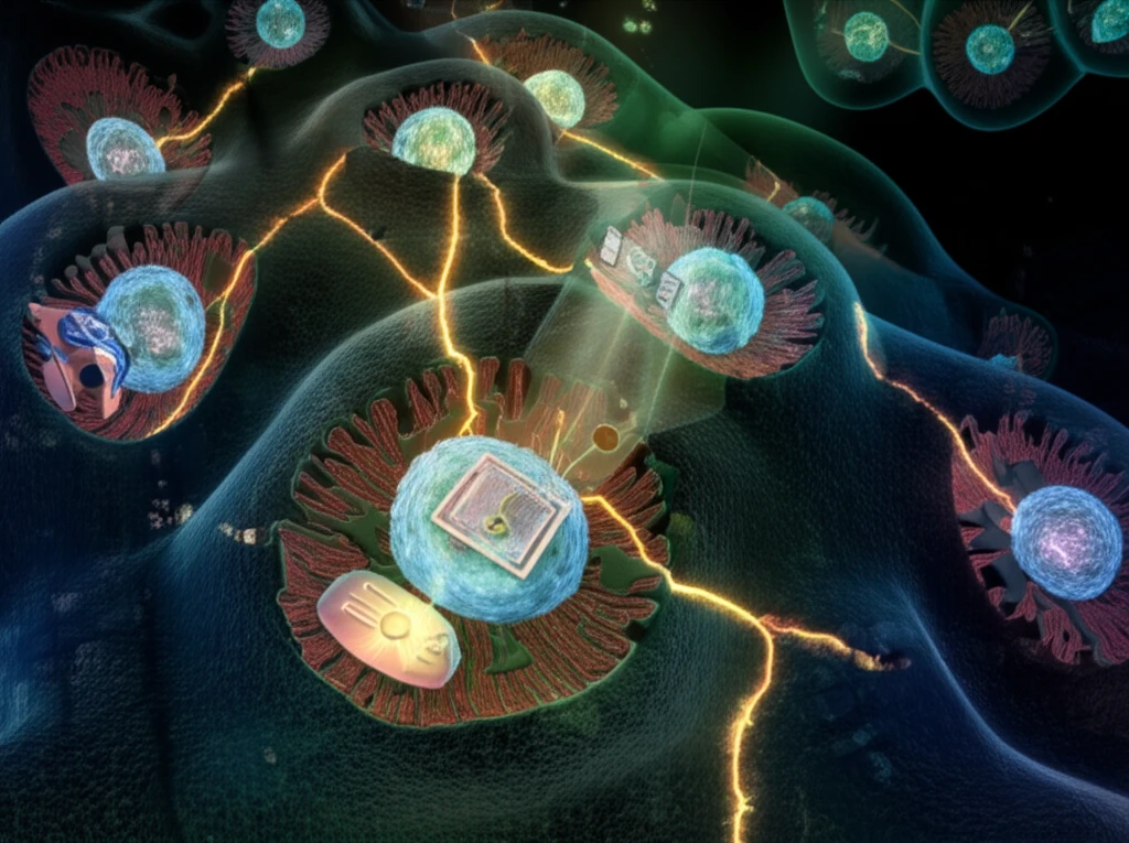
Recent evidence suggests that (P)RR's role as a cell surface receptor may be secondary to more fundamental functions inside the cell. Studies have revealed that (P)RR primarily resides within intracellular organelles, prompting a re-evaluation of its function. In light of this, researchers are directing their focus toward understanding the detailed subcellular distribution of (P)RR and how its location relates to its various functions.
Where is (P)RR Hiding? Unveiling Subcellular Secrets

The (Pro)renin receptor ((P)RR/ATP6ap2) was initially discovered in human mesangial cells and later cloned in 2002. The protein, encoded by the ATP6AP2 gene, consists of 350 amino acids, predicting a mass of around 37 kDa. Its structure includes two hydrophobic domains, suggesting it is a type I transmembrane protein. Researchers have confirmed this structure, showing that (P)RR has an N-terminal signal peptide, an extracellular domain for (pro)renin binding, a single transmembrane domain, and a short cytoplasmic domain.
- A soluble (P)RR of 28-29 kDa, corresponding to the first 278 amino acids.
- A segment including the transmembrane domain and the C-terminal tail.
The Road Ahead: New Directions in (P)RR Research
While the precise functions of (P)RR are still being investigated, its involvement in basic cellular processes like intracellular trafficking is becoming increasingly clear. Future research focusing on how this protein moves within the cell may not only validate known functions but also reveal unexpected roles. Understanding the intricate functions and locations of (P)RR promises to unlock new strategies for treating a range of diseases and improving overall health.
