
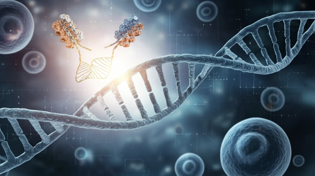
Unlocking the Secrets of DNA: How Genome Editing is Revolutionizing Chromatin Imaging

"Discover how innovative genome editing techniques are transforming our understanding of chromatin dynamics, offering unprecedented insights into gene expression and cellular processes."
For years, scientists have sought to unravel the complexities of chromatin, the intricate structure that packages DNA within our cells. Chromatin dynamics play a crucial role in regulating gene expression, influencing everything from development and differentiation to disease. Traditional methods, such as fluorescence in situ hybridization (FISH), have provided valuable snapshots of DNA distribution, but they lack the ability to capture the dynamic nature of chromatin in living cells.
The limitations of FISH, which requires cell fixation and DNA denaturation, have spurred the development of live imaging techniques. Early approaches involved fusing fluorescent proteins to various chromatin proteins, allowing researchers to visualize overall chromatin structure. However, these methods lacked the specificity needed to target individual genomic loci.
The advent of programmable DNA binding proteins, derived from genome editing technologies, has revolutionized chromatin live imaging. These innovative tools allow scientists to visualize and analyze specific DNA sequences in real-time, opening up new avenues for understanding gene regulation and cellular processes. This article explores these groundbreaking advancements and their potential impact on future research.
Kilobase-Scale Resolution in Living Cells
The latest generation of chromatin imaging operates at remarkable scales: live-cell labeling strategies now allow chromatin to be tracked at single-digit nanometer resolution with millisecond temporal resolution. Super-resolution imaging of single cells has validated many features previously identified by bulk, proximity-based approaches and is challenging existing models of chromatin structure. Live intravital imaging reveals cell-cycle-independent heterogeneity in chromatin compaction across the basal stem cell layer, showing that chromatin gradually rearranges during epidermal stem cell differentiation while transcription of a differentiation-associated gene remains highly dynamic. Deep learning extends the reach of these images: single-cell chromatin images can be binarized into contact matrices that are fed into a variational autoencoder, which projects the high-dimensional contacts into a small set of latent variables that best preserve key features of the original data. Together these advances point toward multiomic single-cell analyses via in situ super-resolution imaging.
From Fixed Tissues to Dynamic Live-Cell Imaging
Understanding chromatin architecture is not limited to visualizing the spatial arrangement of DNA sequences or chromatin compaction; its dynamic behavior is also an essential component, which is why imaging chromatin dynamics in live cells has become a central approach. Single-nucleosome imaging is a promising method because it reveals local chromatin motion that reflects chromatin organization not observed in chemically fixed cells. In living cells, chromatin imaged by several distinct methods displays heterogeneous mobility that depends on its compaction state, subnuclear localization, and ATP-dependent processes. Yet existing tools carry real limitations: super-resolution (SMLM) imaging faces obstacles in image reconstruction, and approaches such as high-resolution diffusion mapping (Hi-D) have been developed to map chromatin motion continuously across the nucleus.
From Nucleosomes to Super-Resolution FISH
Chromatin is the substance within a chromosome consisting of DNA and its associated proteins; the total DNA in a cell is about 5 to 6 feet long and must fit inside the nucleus in an orderly fashion, with DNA molecules first wrapping around histone proteins to form the beads-on-a-string structure called nucleosomes. Early descriptions of this architecture established the basic definition and visual understanding of chromatin that underpins all later work. A major milestone came when multiplexed fluorescence in situ hybridization (FISH) was combined with super-resolution imaging to study chromatin structure. More recent history is marked by a push toward higher-resolution tools: for example, understanding the 3D chromatin conformation of rare cancer stem cells has not been feasible due to the lack of high-resolution chromatin imaging tools.
From Scissors to a Lamp: The Evolution of Chromatin Imaging
The journey from traditional methods to advanced genome editing-based imaging has been marked by significant milestones. Early live imaging techniques relied on fusing fluorescent proteins to general chromatin components. While providing a broad view of chromatin dynamics, these methods lacked the precision to target specific genomic regions. The development of the lacO/LacI system offered improved specificity, but it required the insertion of large DNA sequences into the genome, potentially disrupting normal chromatin structure and function.
- High Specificity: Targets individual genomic loci with precision.
- Real-Time Visualization: Captures the dynamic nature of chromatin in living cells.
- No Disruption: Avoids the insertion of large DNA sequences, preserving normal chromatin structure.
- Versatility: Adaptable for multicolor imaging and signal amplification.
Super-Resolution Probes and Clinical Cohorts
A major technical advance in super-resolution chromatin imaging is the ability to densely label chromatin with oligonucleotide probes that specifically match genomic regions of interest. The approach has already moved into clinical populations: researchers selected a cohort of 57 individuals comprising healthy donors and patients with prediabetes and diabetes, with ages ranging from 25 to 80 years, and performed chromatin imaging of live peripheral blood mononuclear cells (PBMCs) from patients with Type 2 Diabetes Mellitus (T2DM). New methods continue to appear, such as GOLD FISH, presented as a novel chromatin imaging method for studying chromatin structure.
Diffraction Limits and Persistent Obstacles
Despite steady progress, chromatin imaging faces real technical obstacles. The light diffraction limit constrains conventional microscopy, and techniques such as multiphoton and super-resolution microscopy have been highlighted as routes to high-resolution imaging beyond that limit. Scattering-based imaging methods represent another line of technique development, and some of these imaging methods have also been used for imaging human embryos. The continued need to push beyond the diffraction limit underscores how far current standard tools remain from resolving chromatin at the scale where its function is decided.
Fixed Tissue, Live Cells, and Immunostaining Compared
Most studies so far have focused on chromatin structure in cultured cells or in fixed tissue preparations, whereas live imaging approaches offer advantages that these standard preparations cannot. Conventional immunostaining is widely used to label chromatin-associated proteins in many cell types, and the spatial distribution of these proteins provides invaluable information for understanding gene regulation. At a more basic level, comparisons also highlight metabolic differences between chromatin fibers and chromosomes, underscoring that the physical state of DNA changes with the cell cycle. These contrasts — fixed versus live, immunostaining versus imaging-based approaches — frame the trade-offs that shape current experimental choices.
The Future of Chromatin Imaging
Chromatin live imaging with genome editing techniques is poised to revolutionize our understanding of gene regulation and cellular processes. By visualizing the dynamic interactions between DNA sequences and regulatory proteins, researchers can gain insights into the mechanisms underlying development, differentiation, and disease. In the near future, multicolor imaging with multiple guide RNAs will enable the simultaneous visualization of multiple specific chromosomes, offering a powerful alternative to traditional cytogenetic methods. These advances will undoubtedly contribute to new findings in DNA recombination, chromosome rearrangement, epigenetic regulation, and nucleus organization.
Making Dense Chromatin Visible
Directly imaging chromatin within the nucleus is traditionally very difficult because of its dense packaging. This is why experimental model systems matter: Drosophila primary spermatocytes have large nuclei with easily visualized transcriptionally active regions of the Y chromosome known as 'Y loops', making them a valuable system for studying active chromatin. A rich methodological toolkit supports such studies, including protocols for reconstitution of chromatin on solid supports, preparation of positioned mononucleosomes, techniques to study premature chromatin condensation, and the use of comparative genomic hybridization to assess genomic aberrations.
Watching Reprogramming in Real Time
The chromatin regulatory landscape plays a critical role in many disease processes and embryo development, and epigenome sequencing technologies are increasingly profiling that landscape. Live imaging of chromatin in stem cells has so far been performed on undifferentiated cells or during the course of differentiation, but chromatin likely plays essential roles during the reverse process of reprogramming from differentiated, or somatic, cells to induced pluripotent (iPS) cells. Watching chromatin in real time during that reversal would open a frontier that existing imaging studies have not yet reached.
Beyond Bulk Averages
Single-cell imaging validates many of the features previously identified by bulk, proximity-based approaches, and it challenges existing models of chromatin structure. A systemic challenge is that bulk measurements average over large cell populations, so the single-cell picture can overturn assumptions built from population-level data. In situ super-resolution imaging is emerging as a route toward multiomic single-cell analyses, in which one cell can contribute both imaging and molecular data.
Chromatin Signatures for Breast Cancer Detection
Chromatin imaging is emerging as a powerful and straightforward method for assessing cell states and disease stages in ductal carcinoma in situ (DCIS), a precursor of breast cancer, providing a potentially effective biomarker for early detection and treatment planning of breast cancer. Using representation learning on chromatin images alone — without multiplexed staining or high-throughput sequencing — researchers have identified eight morphological cell states and tissue features marking DCIS. These findings suggest that imaging-based signatures could support diagnosis through simpler pipelines that avoid heavy molecular profiling.