
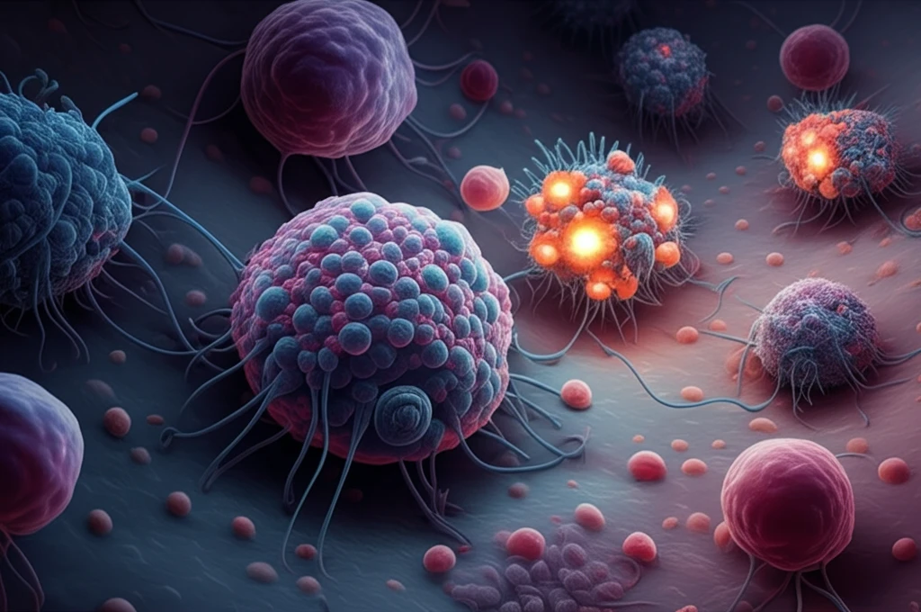
Unlocking the Secrets of Amebiasis: How a Hidden Infection Triggers Inflammation

"Groundbreaking research reveals the crucial roles of caspase-4 and gasdermin D in the inflammatory response to Entamoeba histolytica, paving the way for targeted therapies."
Entamoeba histolytica (Eh), a single-celled parasite, lurks in the shadows, often causing no symptoms at all. Yet, for some, this silent infection transforms into a dangerous invasion, leading to amebiasis, a disease that can trigger severe colitis and life-threatening abscesses in the liver, lungs, or brain. While amebiasis is more prevalent in developing countries with poor sanitation, host factors like genetics and malnutrition also play a critical role in determining who gets sick.
When Eh breaches our innate defenses, the immune system responds with a powerful inflammatory surge designed to eliminate the parasite. However, this response can also harm healthy bystander cells. The delicate balance between protection and damage is orchestrated by a complex interplay of immune cells and signaling molecules.
Macrophages, the sentinels of our immune system, are among the first to encounter Eh. These versatile cells are masters of phagocytosis, engulfing and destroying invaders. But macrophages also produce potent inflammatory cytokines like interleukin (IL)-1β and tumor necrosis factor (TNF)-α, key players in the amebiasis battlefield. This Eh-macrophage interaction is a critical determinant of disease outcome.
A Hidden Infection With Global Reach
Amebiasis is an intestinal illness transmitted when someone eats or drinks something contaminated with a microscopic parasite. It is not common in Canada, occurring more often in parts of the world with poor sanitation, including Mexico, Central America, western South America, southern Asia, and western and southern Africa. Public health agencies such as the CDC maintain dedicated publications, data, and statistics resources on Entamoeba histolytica infection. Both sources agree that contaminated food and water are the central route of transmission.
Diagnosis in the Balance
Standard diagnosis relies on a combination of methods, including dual microscopy, antigen detection, molecular testing, and serologic assays, according to a PLOS Neglected Tropical Diseases review. In many resource-limited settings, diagnosis continues to depend on microscopic identification of cysts and trophozoites in fresh or preserved stool. However, major problems with a wide array of imperfect tests severely limit understanding of the disease's magnitude and epidemiology, with varied and inconsistent application of existing methods in different areas of the world. These diagnostic gaps mean the true burden of amebiasis remains hard to measure.
From First Description to Modern Care
Amebiasis is an intestinal infection caused by the parasite Entamoeba histolytica, a single-celled organism that people acquire by eating or drinking contaminated food or water. The parasite primarily spreads through contaminated food or water. Historically, diagnosis has involved physical examination and attention to travel history in tropical regions. Older clinical accounts also describe complications such as cutaneous amoebiasis, with abscesses, ulcerations, fistulas, or gangrenous inflammation of the skin and subcutis, mostly secondary to active amoebic colitis or amoebic liver abscess.
The Inflammatory Cascade: Caspase-4 and Gasdermin D Take Center Stage
Recent research has shed light on the intricate mechanisms by which Eh triggers inflammation in macrophages. One key virulence factor is the Gal/GalNAc lectin (Gal-lectin), which allows Eh to attach to host cells. This attachment activates caspase-1, an enzyme that initiates the inflammatory response through a complex known as the NLRP3 inflammasome.
- Caspase-4 works independently of some components typically needed for inflammation.
- It teams up with another protein, caspase-1, to boost the release of IL-1β.
- GSDMD is cut apart by caspases, creating pores that let IL-1β escape the cell.
- Gal-Lectin on the parasite help activating both inflammatory proteins.
A Re-Emerging Infection in Developed Countries
Recent research identifies amebiasis, caused by Entamoeba histolytica, as the second leading cause of parasite-related death worldwide. It manifests from asymptomatic carriage to severe clinical conditions such as colitis and liver abscesses, and while it is commonly seen in developing countries, it is re-emerging as a sexually transmitted health problem in developed countries. Ongoing scholarship includes dedicated book-length reviews of the biology and pathogenesis of Entamoeba. Clinical resources consistently describe amebiasis as an infection of the intestines caused by the microscopic parasite E. histolytica.
160 Years Later, Still a Health Problem
Despite roughly 160 years having passed since its first detection, intestinal amoebiasis remains a health problem in developing countries, according to literature indexed in the MalaCards disease database. Amebiasis is an intestinal illness transmitted when someone eats or drinks something contaminated with a microscopic parasite. The long gap between awareness and effective control underscores that knowledge alone has not been enough to eliminate the disease.
Teasing Apart Amebiasis From Other Diseases
Intestinal amebiasis is one of the important differential diagnoses of inflammatory bowel disease in areas where it is highly prevalent. Studies comparing the clinical, endoscopic, and histological features of these disorders had never been done, prompting researchers to undertake this comparison. Research on the immune response in amebiasis and immune evasion by E. histolytica continues to clarify how the parasite interacts with host defenses. For clinicians, side-by-side comparison tools for amebiasis medications are available, covering coverage, dosing, adverse reactions, and savings programs.
A Novel Role for Caspase-4: Sensing and Amplifying the Inflammatory Signal
These findings reveal a novel role for caspase-4 as a sensor molecule that amplifies pro-inflammatory responses when macrophages encounter Eh. Targeting this pathway could offer a new approach to managing the damaging inflammation associated with amebiasis, potentially leading to more effective therapies.
Expert Consensus and Clinical Pearls
Expert synthesis describes amebiasis as an infection primarily caused by the protozoan parasite Entamoeba histolytica, transmitted mainly through the fecal-oral route but also spread sexually via oral-anal contact. Specialist physicians rated as elite in treating amebiasis, such as Kris Chadee in Calgary, Canada, also work on related conditions including giardia infection, colitis, and viral gastroenteritis. Historically, radiologists have found unique value in the therapeutic test with metronidazole for arriving at a final, conclusive roentgen diagnosis of intestinal amebiasis.
New Routes of Research and Investment
Future directions for control focus on sexually transmitted amebiasis, a re-emerging health problem in developed countries, with dedicated research outlining strategies for managing this form of infection. Market analyses project a promising outlook for the amebiasis drugs sector, citing increasing investment in research and development and growing awareness about the disease. Together, these outlooks point to both clinical and commercial momentum behind addressing the infection's changing epidemiology.
A Parasite Unique to Humans
Amebiasis is caused by a one-celled parasite, Entamoeba histolytica, which is found only in human beings. Transmission occurs via the fecal-oral route, either directly through person-to-person contact such as diaper changing and sexual practices, or when a person ingests cysts through contaminated food or water. Certain groups face higher risk, including men who have sex with other men and people with compromised immune systems or other health conditions. Because the parasite lives only in humans, its control depends on interrupting person-to-person spread as much as on sanitation.
When the Parasite Turns on Its Host
In many cases, the parasite that causes amebiasis lives in a person's large intestine without causing any symptoms. When disease does occur, the initial site of amebic infection is the cecum and colon after E. histolytica excysts in the small bowel, with trophozoite attachment to the colonic mucosa followed by mucosal invasion that produces superficial and deep colonic ulcerations. The infection can be especially severe in the most vulnerable patients: one reported case involved a five-month-old girl who exhibited vomiting, refusal to feed, abdominal distension, and mucoid, bloody stools. Clinically, amebiasis behaves as a chronic, relapsing colitis that can also carry extraintestinal manifestations.