Glowing brain representing posterior reversible encephalopathy syndrome (PRES).

Unlocking the Mystery of PRES: What You Need to Know About This Reversible Brain Condition

"Atypical cases reveal critical insights into posterior reversible encephalopathy syndrome, emphasizing the importance of early diagnosis and management."


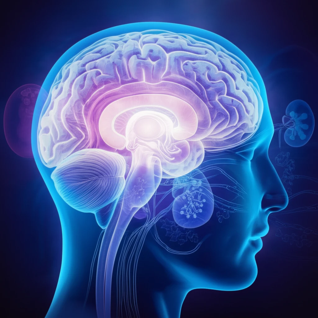
Posterior reversible encephalopathy syndrome (PRES) is a condition that can affect the brain, typically causing temporary problems. Usually, PRES involves the back part of the brain. It's known for being reversible, meaning patients can recover well with the right treatment. Understanding this condition is vital for early diagnosis and effective management.

PRES is often linked to conditions like severe high blood pressure, kidney failure, autoimmune disorders, certain medications, and eclampsia. Common signs include confusion, seizures, headaches, and vision problems. Doctors use MRI scans to look for swelling in the brain, particularly in the white matter, which is often seen in the posterior regions.

While PRES usually affects the back of the brain, a recent case highlights an unusual presentation involving the temporal pole. This case emphasizes that PRES can occur in different ways, and recognizing these variations is crucial for proper diagnosis and treatment. The involvement of the deep white matter, including the temporal pole, is rarely seen in ischemic stroke, making it a potential sign of PRES.

Atypical PRES Case: Deep White Matter Involvement

Glowing brain representing posterior reversible encephalopathy syndrome (PRES).

A 55-year-old man was admitted to the emergency department with mild confusion. Over the previous three months, he had been making mistakes at work and feeling fatigued. On the day of admission, he couldn't stand up on his own. He had a history of untreated high blood pressure (170/120 mmHg) for three years, along with alcohol abuse, gastric ulcers, and a cleft lip palate. His diet was poor, and he smoked 40 cigarettes daily for 25 years.

Upon examination, his blood pressure was alarmingly high at 269/189 mmHg, and his pulse rate was 105 beats per minute. He was slightly disoriented, had trouble perceiving time, and communication was difficult. A neurological exam revealed only abnormal eye movements. Other motor and sensory functions were normal.

Laboratory tests showed:
  • Hemoglobin: 12.8 g/dL
  • Leukocyte Count: 14,050/mm²
  • Platelet Count: 140,000/mm²
  • Serum Creatinine: 8.43 mg/dL
  • Urea Nitrogen: 106 mg/dL
  • Mild hypokalemia (potassium: 3.0 mEq/L)
Based on these findings, he was diagnosed with acute hypertensive nephrosclerosis (kidney damage due to high blood pressure) and acute renal failure. Brain CT scans showed swelling in the brainstem, cerebellum, and deep white matter. MRI scans confirmed vasogenic edema (fluid accumulation) in these areas. Despite the severe MRI findings, his symptoms were relatively mild.

Key Takeaways: Recognizing PRES and Its Variations

This case highlights the importance of considering PRES in patients with severe hypertension and neurological symptoms, even when the presentation is atypical. The involvement of the deep white matter, especially the temporal pole, is a less common finding but should raise suspicion for PRES.

Early diagnosis and treatment, including blood pressure control and hemodialysis, can lead to significant clinical and radiological improvement. Recognizing the varied presentations of PRES is crucial for timely intervention and better patient outcomes.

While the pathophysiology of PRES is not fully understood, rapidly developing hypertension is believed to disrupt cerebral blood flow autoregulation, leading to vasogenic edema. Further research is needed to better understand the mechanisms underlying PRES and identify potential risk factors.

About this Article -

This article was crafted using a human-AI hybrid and collaborative approach. AI assisted our team with initial drafting, research insights, identifying key questions, and image generation. Our human editors guided topic selection, defined the angle, structured the content, ensured factual accuracy and relevance, refined the tone, and conducted thorough editing to deliver helpful, high-quality information.See our About page for more information.

This article is based on research published under:

DOI-LINK: 10.2169/internalmedicine.55.7324, Alternate LINK

Title: Posterior Reversible Encephalopathy Syndrome With Extensive Deep White Matter Lesions Including The Temporal Pole

Subject: General Medicine

Journal: Internal Medicine

Publisher: Japanese Society of Internal Medicine

Authors: Junichiro Ohira, Nobuyuki Mori, Shunsuke Kajikawa, Takeshi Nakamura, Tetsuya Arisato, Makio Takahashi

Published: 2016-01-01

Everything You Need To Know

1

What is posterior reversible encephalopathy syndrome (PRES)?

The condition is called posterior reversible encephalopathy syndrome (PRES). It's a brain condition characterized by temporary neurological issues. It primarily affects the back portion of the brain and is often reversible with appropriate treatment. Understanding PRES is crucial for timely diagnosis and effective treatment strategies.

2

What are the typical causes of PRES?

PRES is often associated with several underlying conditions, including severe high blood pressure, kidney failure, autoimmune disorders, certain medications, and eclampsia. In the context of the case, the patient's untreated high blood pressure of 170/120 mmHg for three years, escalating to 269/189 mmHg upon admission, was a significant contributing factor. Other factors, such as kidney damage and alcohol abuse, likely contributed to the development of PRES.

3

What are the common symptoms of PRES?

Common signs of PRES include confusion, seizures, headaches, and vision problems. The case described a 55-year-old man who presented with mild confusion, mistakes at work, and fatigue over several months. Upon admission, he showed disorientation, difficulty with communication, and abnormal eye movements. These symptoms are critical indicators prompting medical investigation using MRI scans.

4

How is PRES typically diagnosed, and what are some atypical presentations?

In typical cases of PRES, doctors use MRI scans to detect swelling in the brain, specifically in the white matter, particularly in the posterior regions. However, the case mentioned showed an atypical presentation. In this instance, there was involvement of the deep white matter, including the temporal pole, an uncommon finding. This highlights the importance of considering different presentations of PRES for accurate diagnosis and treatment.

5

Why is it important to detect and manage PRES early?

Early detection and proper management of PRES can lead to a full recovery. The patient's diagnosis of acute hypertensive nephrosclerosis (kidney damage due to high blood pressure) and acute renal failure, along with MRI findings of vasogenic edema, highlight the importance of managing the underlying causes. The treatment typically focuses on controlling blood pressure and addressing the underlying conditions contributing to PRES. Prompt intervention is key to reversing the effects of the condition and achieving a favorable outcome.

Newsletter Subscribe

Subscribe to get the latest articles and insights directly in your inbox.