
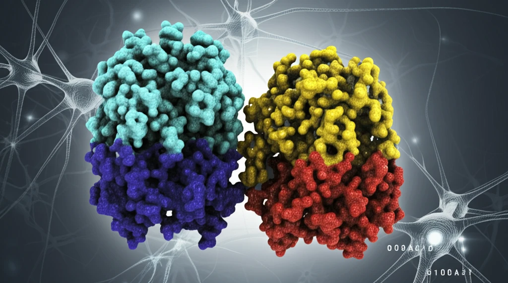
Unlock the Secrets of Cellular Stress: How STIP1 and S100A1 Interaction Could Revolutionize Brain Health

"Delve into the molecular dance between STIP1 and S100A1 and discover how their intricate relationship offers a new path for understanding and treating neurodegenerative diseases, using the Power of Proteins."
Our bodies are constantly under siege from various stressors, both internal and external. At the cellular level, these stresses can disrupt normal functions, leading to a cascade of problems, particularly in sensitive tissues like the brain. Central to managing this cellular stress are two proteins: Stress-Inducible Phosphoprotein 1 (STIP1) and S100A1. These proteins are involved in a complex interaction that scientists are now beginning to understand, and it's this understanding that could pave the way for revolutionary treatments for neurodegenerative diseases.
STIP1 acts as a crucial coordinator in the cell, helping other proteins, known as heat shock proteins (Hsp70 and Hsp90), to fold correctly. This folding process is essential for proteins to function properly; when it goes awry, it can lead to the accumulation of misfolded proteins, a hallmark of diseases like Alzheimer's. Meanwhile, S100A1, a calcium-binding protein, has been found to influence how STIP1 interacts with these heat shock proteins. The relationship between STIP1 and S100A1 is like a delicate dance, where each protein's actions affect the other, and understanding this dance is key to maintaining cellular health.
Recent research has illuminated the molecular basis for the interaction between STIP1 and S100A1, revealing how they bind and influence each other's functions. These findings not only deepen our knowledge of cellular stress mechanisms but also highlight potential therapeutic targets for neurological disorders. By manipulating the interaction between these proteins, we might be able to prevent or even reverse the damage caused by misfolded proteins in the brain.
Molecular Basis of the S100A1–STIP1 Interaction
The interaction between S100A1 and STIP1 has been characterized at the molecular level using isothermal titration calorimetry (ITC). ITC thermograms revealed a high-affinity binding event between S100A1 and full-length STIP1, providing thermodynamic data on the strength and nature of their association. This biophysical evidence establishes that these two proteins form a stable complex under physiological conditions, which is a prerequisite for any functional role in cellular stress pathways. Understanding the binding energetics is an essential first step toward exploring downstream biological consequences.
Characterization Techniques and Constraints
Isothermal titration calorimetry is a standard technique used to quantify the thermodynamic parameters of protein–protein interactions, including binding affinity, stoichiometry, enthalpy, and entropy. While ITC provides label-free, solution-phase data that closely mimics physiological conditions, it requires relatively large quantities of purified protein and cannot easily resolve conformational changes occurring during binding. Complementary methods such as surface plasmon resonance, co-immunoprecipitation, and fluorescence-based assays are often needed to validate and extend ITC findings. Each technique carries its own assumptions and limitations, meaning no single method can fully characterize a protein interaction in isolation.
Tracing the Discovery Timeline
The S100 protein family was first identified in the 1960s as a brain-enriched, soluble protein fraction, and subsequent decades revealed its involvement in calcium signaling and neurodegeneration. STIP1, also known as stress-induced phosphoprotein 1 or HOP (Hsp70/Hsp90-organizing protein), was characterized as a co-chaperone that bridges the Hsp70 and Hsp90 chaperone systems. The recognition that S100A1 and STIP1 physically interact emerged from proteomic and biophysical studies investigating stress-response networks in neural tissue. These foundational discoveries laid the groundwork for investigating how the S100A1–STIP1 axis modulates cellular stress responses relevant to brain health.
The Molecular Basis of STIP1 and S100A1 Interaction
The study reveals that S100A1 binds to STIP1 through specific regions called tetratricopeptide repeat (TPR) domains. STIP1 has three TPR domains (TPR1, TPR2A, and TPR2B), each capable of binding to S100A1. Interestingly, S100A1 doesn't bind to STIP1 as a single unit but rather as a dimer—two S100A1 molecules joined together. Isothermal titration calorimetry, a technique used to measure the heat changes associated with binding events, showed that each TPR domain binds a single S100A1 dimer, but with varying strength. The TPR2B domain exhibited the highest affinity for S100A1, suggesting it plays a critical role in the interaction.
- S100A1 binds to STIP1's TPR domains as a dimer.
- Calcium binding to S100A1 is essential for the interaction.
- TPR2B domain shows the highest affinity.
- The binding involves a hydrophobic cleft exposed by calcium.
Emerging Findings on Stress-Response Protein Networks
Recent studies have begun to map the broader interactome of S100A1, identifying multiple binding partners implicated in neuroprotection and neurodegeneration. Research into stress-induced phosphoprotein 1 (STIP1) has highlighted its role as a hub connecting molecular chaperone pathways, with implications for protein homeostasis in the brain. Investigations combining biophysical binding data with cell-based assays are increasingly being used to assess the functional consequences of S100A1–STIP1 complex formation. These converging lines of evidence suggest that disruption of this interaction could contribute to the protein misfolding cascades characteristic of neurodegenerative disease.
Challenges in Translating S100 Biomarker Data to Clinical Practice
S100 proteins, including S100A1 and S100B, have been proposed as early biomarkers for traumatic brain injury, stroke, and brain hypoxia. However, clinical deployment has been hampered by the fact that S100 is not exclusively produced in the central nervous system—it is also released from melanoma tumors and other extracerebral sources, complicating diagnostic specificity. Elevated serum S100 levels must therefore be interpreted with caution, as they may reflect peripheral tissue damage rather than brain-specific injury. This limitation underscores the need for multi-marker panels and contextual clinical assessment rather than reliance on S100 measurement alone.
S100A1 in the Context of Immune Cell Signaling
Comparative transcriptomic studies of macrophage polarization states have revealed distinct gene signatures associated with classically activated (M1) versus alternatively activated (M2) phenotypes. Among the upregulated genes in M1-polarized macrophages are several chemokines, including Ccl7, Ccl17, Ccl22, and Ccl24, which coordinate inflammatory recruitment. S100-family proteins, including S100A1, are known to be expressed in activated immune cells and can modulate inflammatory signaling cascades. Placing the S100A1–STIP1 interaction within this broader immunological gene-expression landscape suggests a potential role in linking calcium-dependent immune activation to chaperone-mediated protein quality control.
Implications for Future Therapies
The detailed understanding of the STIP1-S100A1 interaction offers promising new directions for therapies targeting Alzheimer's and other neurological disorders. By understanding the molecular basis, researchers can design molecules that specifically interfere with or enhance this interaction, potentially preventing protein misfolding and reducing the toxic effects on brain cells. The findings suggest that modulating the interaction between STIP1 and S100A1 could be a key strategy in combating neurodegenerative diseases.
Integrating Biophysical and Systems-Level Perspectives
The convergence of biophysical binding studies and systems-level analyses offers a more complete picture of how S100A1 and STIP1 function together. While ITC provides the quantitative thermodynamic foundation, network-level analyses help identify the downstream pathways that this interaction may regulate. Expert interpretation of these data suggests that the S100A1–STIP1 complex sits at a critical node linking calcium signaling, chaperone activity, and stress adaptation in neural cells. However, translating these molecular insights into mechanistic models of brain disease requires further interdisciplinary synthesis.
Unresolved Questions and Path Forward
Key open questions include whether the S100A1–STIP1 interaction is dynamically regulated by calcium fluctuations during acute cellular stress, and whether pharmacological stabilization of this complex could be neuroprotective. Structural biology approaches such as cryo-electron microscopy may resolve the atomic-level architecture of the complex and identify druggable interfaces. In vivo studies using conditional knockout models will be needed to establish causality between this protein interaction and neurological outcomes. The field is positioned to move from correlative binding data toward functional and translational studies in the coming years.
Translational Barriers in Neurodegeneration Research
The path from identifying a protein–protein interaction to developing a therapeutic intervention is long and fraught with systemic challenges, including the complexity of the blood–brain barrier, species differences in protein expression, and the heterogeneity of neurodegenerative disease pathology. Biomarker studies have shown that even well-characterized markers like S100B face significant hurdles in clinical translation due to issues of specificity and standardization across laboratories. Regulatory and funding landscapes further shape which research avenues receive sustained support. Addressing these broader contextual factors is essential for any laboratory finding—such as the S100A1–STIP1 interaction—to ultimately benefit patients.
From Bench to Bedside: The Human Stakes
Ultimately, research into the S100A1–STIP1 interaction is motivated by the human toll of neurodegenerative diseases, which affect millions worldwide and currently lack disease-modifying therapies. Naturalistic observation and case-study methodologies remain valuable tools for understanding how molecular findings manifest in real-world clinical presentations. The gap between mechanistic laboratory discoveries and patient outcomes underscores the importance of translational research frameworks that incorporate both molecular and clinical perspectives. Bridging this gap requires sustained collaboration between basic scientists, clinicians, and patients.