Comparison of ultrasound and MRI for muscle volume measurement.

Ultrasound vs. MRI: Which is the Best Way to Measure Muscle Volume?

"A new study compares 3D ultrasound to MRI for measuring gastrocnemius muscle volume in adults and children with and without cerebral palsy, revealing surprising insights for athletes and patients."


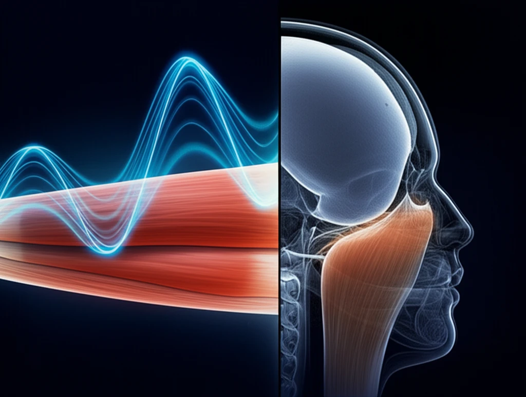
Understanding muscle volume is crucial for assessing muscle health, tracking adaptations to training, and diagnosing various conditions. Historically, MRI has been considered the gold standard for measuring muscle volume due to its high-resolution imaging capabilities. However, MRI has limitations, including high costs, long scanning times, and contraindications for individuals with metal implants or epilepsy. These limitations have spurred the search for alternative, more accessible methods for assessing muscle volume.

Ultrasound, particularly freehand 3D ultrasound (3DUS), has emerged as a promising alternative. 3DUS offers several advantages over MRI, including lower cost, portability, and shorter scanning times. However, questions remain about the accuracy and reliability of 3DUS compared to MRI, especially in diverse populations such as children and individuals with neuromuscular conditions like cerebral palsy.

A recent study published in 2023 directly compared 3DUS to MRI for measuring the gastrocnemius muscle volume in adults and children, with and without cerebral palsy. The study aimed to evaluate the validity of 3DUS and explore its potential as a practical tool for assessing muscle health in various clinical and research settings.

How Accurate is 3D Ultrasound Compared to MRI?

Comparison of ultrasound and MRI for muscle volume measurement.

The study meticulously compared muscle volume measurements obtained from 3DUS and MRI. The results indicated a high level of agreement between the two methods. Specifically, the mean difference in muscle volume measurements between MRI and 3DUS was minimal, with strong correlation coefficients, suggesting that 3DUS can accurately measure muscle volume. Furthermore, digitizing errors for 3DUS were minimal, demonstrating the reliability of the technique.

The research sheds light on the precision of ultrasound in assessing muscle characteristics, making it a viable option for settings where MRI is not feasible. For those monitoring their athletic performance or undergoing rehabilitation, this data provides a reassuring perspective on the reliability of ultrasound technology.

  • Accuracy: High agreement between 3DUS and MRI measurements.
  • Reliability: Minimal digitizing errors for 3DUS.
  • Versatility: Accurate in adults, typically developing children, and children with cerebral palsy.
The study highlights that 3DUS is a valid method for muscle volume measurement across different age groups and populations, offering a practical alternative to MRI.

The Future of Muscle Assessment: Broader Implications

The findings of this study have significant implications for clinical practice and sports medicine. 3DUS offers a cost-effective, portable, and time-efficient alternative to MRI for assessing muscle volume. This is particularly valuable in settings where MRI is not readily available or feasible. The ability to accurately measure muscle volume with 3DUS can aid in the diagnosis and monitoring of various conditions, as well as in tracking training adaptations in athletes. Further research is needed to explore the use of 3DUS in different muscle groups and populations.

About this Article -

This article was crafted using a human-AI hybrid and collaborative approach. AI assisted our team with initial drafting, research insights, identifying key questions, and image generation. Our human editors guided topic selection, defined the angle, structured the content, ensured factual accuracy and relevance, refined the tone, and conducted thorough editing to deliver helpful, high-quality information.See our About page for more information.

This article is based on research published under:

DOI-LINK: 10.1002/ca.23314, Alternate LINK

Title: A Comparison Of 3D Ultrasound To Mri For The Measurement And Estimation Of Gastrocnemius Muscle Volume In Adults And Young People With And Without Cerebral Palsy

Subject: General Medicine

Journal: Clinical Anatomy

Publisher: Wiley

Authors: Marika Noorkoiv, Nicola Theis, Grace Lavelle

Published: 2019-01-09

Everything You Need To Know

1

Why has MRI traditionally been considered the gold standard for measuring muscle volume?

MRI has been considered the gold standard for measuring muscle volume due to its high-resolution imaging capabilities. This allows for detailed visualization and precise measurement of muscle tissue. However, MRI has limitations, including high costs, long scanning times, and contraindications for individuals with metal implants or epilepsy. These limitations have led to the exploration of alternative methods like 3D ultrasound (3DUS). While MRI provides excellent detail, its accessibility and suitability for all patients are factors that drive the need for alternative assessment tools.

2

What are the primary advantages of using freehand 3D ultrasound (3DUS) over MRI for measuring muscle volume?

Freehand 3D ultrasound (3DUS) offers several advantages over MRI for measuring muscle volume. These include lower cost, portability, and shorter scanning times. 3DUS is more accessible, especially in settings where MRI is not readily available or feasible. Furthermore, 3DUS does not have the same contraindications as MRI, making it suitable for a broader range of individuals, including those with metal implants or epilepsy. The speed and cost-effectiveness of 3DUS make it a practical alternative for routine muscle assessments and monitoring.

3

How accurate is 3D ultrasound (3DUS) in measuring muscle volume compared to MRI, according to recent studies?

Recent studies indicate a high level of agreement between 3D ultrasound (3DUS) and MRI in measuring muscle volume. The mean difference in muscle volume measurements between MRI and 3DUS is minimal, with strong correlation coefficients. This suggests that 3DUS can accurately measure muscle volume, making it a viable alternative to MRI. The digitizing errors for 3DUS are also minimal, demonstrating the reliability of the technique. This accuracy makes 3DUS a valuable tool for assessing muscle health in various clinical and research settings.

4

What are the broader implications of using 3D ultrasound (3DUS) for muscle assessment in clinical practice and sports medicine?

The use of 3D ultrasound (3DUS) has significant implications for clinical practice and sports medicine. 3DUS offers a cost-effective, portable, and time-efficient alternative to MRI for assessing muscle volume. This is particularly valuable in settings where MRI is not readily available or feasible. The ability to accurately measure muscle volume with 3DUS can aid in the diagnosis and monitoring of various conditions, as well as in tracking training adaptations in athletes. The accessibility and ease of use of 3DUS can improve the efficiency of muscle assessments and expand access to care.

5

In what specific populations has 3D ultrasound (3DUS) been proven to be a valid method for muscle volume measurement?

3D ultrasound (3DUS) has been proven to be a valid method for muscle volume measurement across different age groups and populations, including adults, typically developing children, and children with cerebral palsy. Studies have shown that 3DUS provides accurate and reliable measurements of muscle volume in these diverse groups, making it a versatile tool for assessing muscle health in various clinical and research settings. The ability to use 3DUS in these different populations highlights its potential as a practical and accessible alternative to MRI for a wide range of individuals.

Newsletter Subscribe

Subscribe to get the latest articles and insights directly in your inbox.