3D rendering of a cancer cell nucleus showing the complex organization of DNA and key nuclear components.

The 3D Cancer Nucleus: A New Dimension in Understanding and Treating Cancer

"Delving into the spatial organization of the cancer cell genome could revolutionize cancer diagnostics and treatment."


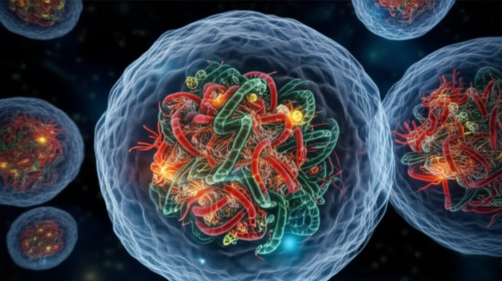
For over a century, scientists have been exploring the intricate world within the cell's nucleus, particularly how the organization of chromosomes changes in cancer. Groundbreaking early observations by Hansemann and Boveri laid the foundation for our current understanding of the cancer cell. Their work highlighted the abnormal structure and behavior of nuclei and chromosomes in cancerous cells, sparking decades of research into the underlying causes and potential therapeutic targets.

Today, modern molecular and imaging techniques are providing an unprecedented look at the spatial arrangement of the cancer cell's genome. This deeper understanding is revealing that the structural order of the nucleus isn't just a static feature—it's a dynamic element that can serve as a biomarker for cancer.

This article explores the exciting advancements in understanding the 3D organization of the cancer nucleus, from its historical roots to its potential applications in cancer diagnosis and personalized medicine. We'll delve into the key discoveries, technological breakthroughs, and future directions that promise to reshape our approach to cancer care.

Unlocking the Secrets of the 3D Genome: Key Discoveries and Technologies

3D rendering of a cancer cell nucleus showing the complex organization of DNA and key nuclear components.

Research into the spatial organization of the cancer nucleus has been accelerated by advancements in several key areas:

  • Fluorescent in situ hybridization (FISH): This technique allows scientists to visualize the location of specific DNA sequences within the nucleus, providing insights into chromosome positioning and organization in 3D.
  • Chromosome conformation capture technologies: Methods like Hi-C reveal how different regions of the genome interact with each other, mapping the complex network of contacts within the nucleus.
  • Super-resolution microscopy: Breaking the diffraction limit of light, these advanced imaging techniques provide unprecedented detail of nuclear architecture, revealing the organization of DNA and other components at the nanoscale.

These technologies have enabled researchers to identify several key features of the 3D cancer nucleus:

The Future of Cancer Care: Targeting the 3D Nucleus

The research paints a promising picture: By understanding the intricacies of the 3D cancer nucleus, we can develop new diagnostic tools, personalize treatment strategies, and potentially even target the nuclear architecture itself to disrupt cancer development.

While significant progress has been made, further research is crucial. Large-scale studies are needed to validate these findings and translate them into clinical applications. Multi-center collaborations and clinical trials will be essential to ensure that these innovative approaches benefit all cancer patients.

The journey into the 3D cancer nucleus is ongoing, but the potential rewards are immense. By continuing to explore this new dimension of cancer biology, we can unlock new possibilities for prevention, diagnosis, and treatment, bringing hope to millions affected by this disease.

About this Article -

This article was crafted using a human-AI hybrid and collaborative approach. AI assisted our team with initial drafting, research insights, identifying key questions, and image generation. Our human editors guided topic selection, defined the angle, structured the content, ensured factual accuracy and relevance, refined the tone, and conducted thorough editing to deliver helpful, high-quality information.See our About page for more information.

Everything You Need To Know

1

Why is the spatial organization of the cancer cell genome so important?

The spatial arrangement of the cancer cell genome within the 3D cancer nucleus is crucial because it influences gene expression, DNA replication, and DNA repair. Disruptions in this organization can lead to abnormal cellular behavior and contribute to cancer development. By understanding how the genome is organized in three dimensions, researchers can identify new biomarkers for cancer diagnosis and develop therapies that target specific structural features of the nucleus to disrupt cancer development.

2

What are Fluorescent in situ hybridization, Chromosome conformation capture technologies and super-resolution microscopy?

Fluorescent in situ hybridization, or FISH, allows researchers to visualize the location of specific DNA sequences within the nucleus, helping them understand chromosome positioning and organization. Chromosome conformation capture technologies, like Hi-C, map the complex network of interactions between different regions of the genome. Finally, super-resolution microscopy provides detailed images of the nuclear architecture at the nanoscale, revealing the organization of DNA and other components.

3

How did Hansemann and Boveri contribute to our understanding of the cancer cell?

Hansemann and Boveri's early work highlighted the abnormal structure and behavior of nuclei and chromosomes in cancerous cells. Their observations suggested that these abnormalities were not random, but rather indicative of underlying genetic and cellular changes driving cancer development. This laid the foundation for future research into the causes of cancer, which led to potential therapeutic interventions that focused on the nucleus.

4

How can targeting the 3D cancer nucleus revolutionize cancer care?

Targeting the 3D cancer nucleus could lead to new diagnostic tools by identifying unique spatial biomarkers specific to cancer cells. Personalized treatment strategies could be developed based on the individual patient's nuclear architecture. There's also potential to directly target the nuclear architecture to disrupt cancer development. Understanding the 3D nucleus can help detect the disease earlier, and tailor treatments.

5

What are the limits of the research, and what relevant technologies are omitted?

While the research has focused on FISH, Hi-C, and super-resolution microscopy, it would be interesting to know more about the computational methods used to analyze the vast amounts of data generated by these technologies. These methods are essential for interpreting the complex interactions within the nucleus. A deeper discussion of clinical trials using therapeutic strategies that target the 3D cancer nucleus would provide insight into practical applications of these research areas.

Newsletter Subscribe

Subscribe to get the latest articles and insights directly in your inbox.