Spinal Sarcoidosis: Spotting the Tricky Signs and What It Means for You
"Unraveling the complexities of spinal sarcoidosis: From early detection to innovative imaging techniques, empowering you with knowledge for proactive health management."
Spinal sarcoidosis, a rare manifestation of sarcoidosis, involves the spine and can easily be mistaken for other neurological conditions. This article sheds light on this often-overlooked condition, providing insights into early diagnosis, imaging techniques, and treatment responses.
Sarcoidosis, characterized by inflammation, primarily affects the lungs, skin, and lymph nodes. When it extends to the central nervous system (CNS), including the spinal cord, it's termed neurosarcoidosis. Spinal sarcoidosis, either occurring with or independently of CNS involvement, presents diagnostic challenges due to its non-specific symptoms.
This article aims to translate complex research into understandable information, focusing on the early detection of spinal sarcoidosis through magnetic resonance imaging (MRI), how to interpret imaging findings, and what these findings mean for managing the condition. Learn about innovative techniques like diffusion-weighted imaging that offer new insights into disease activity and tissue damage.
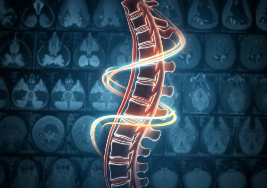
Decoding Spinal Sarcoidosis: What the Images Reveal
Magnetic Resonance Imaging (MRI) is crucial for detecting spinal sarcoidosis, often revealing:
- Leptomeningeal enhancement (inflammation of the membranes surrounding the brain and spinal cord): 61%
- Pachymeningeal enhancement (inflammation of the outer membrane of the brain and spinal cord): 23%
- Intramedullary enhancing lesions (lesions within the spinal cord): 38%
- Bony involvement: 15%
The Future of Spinal Sarcoidosis Detection and Management
Spinal sarcoidosis, once considered rare, is increasingly recognized due to advancements in MRI technology. Characteristic imaging patterns, especially dorsal surface involvement, aid in diagnosis, although they aren't definitive.
Quantitative diffusion studies, like diffusion-weighted imaging (DWI), offer a new way to assess disease activity and tissue damage. These techniques measure the apparent diffusion coefficient (ADC), which can show changes in the spinal cord tissue. For example, intramedullary lesions often exhibit increased ADC values, indicating vasogenic edema and inflammation.
While imaging findings are essential, clinical symptoms don't always mirror MRI results. Close monitoring and collaboration between neurologists and radiologists are crucial for tailoring treatment plans and improving outcomes in spinal sarcoidosis.