Liver with puzzle pieces representing the diagnostic challenge of liver lesions

Rare Liver Cancer Mimics: When Imaging Isn't Enough

"Contrast-enhanced ultrasound can be a powerful tool, but sometimes a liver biopsy is the only way to get a definitive diagnosis."


When it comes to liver health, early detection is key. Ultrasound is often the first line of defense, helping doctors spot potential problems quickly and non-invasively. Contrast-enhanced ultrasound (CEUS) takes it a step further, offering a clearer picture of liver lesions and helping to differentiate between cancerous and non-cancerous growths.

However, even the most sophisticated imaging has its limitations. Primary hepatic lymphoma (PHL), a rare cancer that starts in the liver, can sometimes mimic other liver conditions on imaging, making accurate diagnosis a challenge. This is where a liver biopsy becomes essential.

This article explores two cases where CEUS and CT scans weren't enough to distinguish PHL from more common liver lesions like hepatocellular carcinoma (HCC). We'll delve into why a liver biopsy was necessary for a definitive diagnosis and highlight the importance of considering rare possibilities when evaluating liver abnormalities.

The Challenge of Diagnosing Primary Hepatic Lymphoma

Liver with puzzle pieces representing the diagnostic challenge of liver lesions

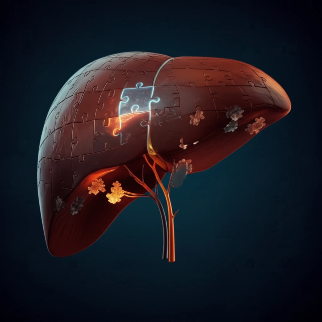
Primary hepatic lymphoma (PHL) is an exceedingly rare cancer, often overshadowed by more prevalent liver conditions. Its subtle presentation can easily lead to misdiagnosis, making it a diagnostic puzzle for clinicians.

In a study following two cases of patients with hepatitis B, ultrasound results showed similar characteristics to HCC (hepatocellular carcinoma). Here's what happened:

  • Both patients had a history of hepatitis B virus (HBV) infection.
  • Ultrasound revealed hypoechoic liver lesions with irregular margins.
  • CEUS showed hyperenhancement in the arterial phase and hypoenhancement in the portal and late phases, mimicking HCC.
  • Contrast-enhanced CT scans also suggested HCC.
Despite the imaging results pointing towards HCC, the doctors suspected that something else was afoot. The absence of cirrhosis, a common risk factor for HCC, prompted them to perform liver biopsies. The biopsies revealed the true diagnosis: marginal zone B-cell lymphoma of the mucosa-associated lymphoid tissue (MALT) type, a subtype of PHL.

The Importance of Considering Rare Possibilities

These cases highlight a crucial lesson: even when imaging strongly suggests a particular diagnosis, it's important to consider less common possibilities, especially when clinical factors don't quite align.

While CEUS is a valuable tool for evaluating liver lesions, it's not foolproof. PHL can mimic HCC on imaging, leading to potential misdiagnosis and delayed treatment. In patients with risk factors for lymphoproliferative disorders, such as chronic HBV infection, PHL should be on the list of differential diagnoses.

When imaging results are inconclusive or don't match the clinical picture, a liver biopsy remains the gold standard for definitive diagnosis. It's the key to unlocking the mystery and ensuring patients receive the right treatment, especially for rare conditions like primary hepatic lymphoma.

About this Article -

This article was crafted using a human-AI hybrid and collaborative approach. AI assisted our team with initial drafting, research insights, identifying key questions, and image generation. Our human editors guided topic selection, defined the angle, structured the content, ensured factual accuracy and relevance, refined the tone, and conducted thorough editing to deliver helpful, high-quality information.See our About page for more information.

Everything You Need To Know

1

What is the role of ultrasound and contrast-enhanced ultrasound (CEUS) in evaluating liver health?

Ultrasound is a non-invasive imaging technique that's often used to initially assess the liver for potential problems. Contrast-enhanced ultrasound (CEUS) is a more advanced form of ultrasound that uses contrast agents to enhance the images, providing a clearer picture of liver lesions. However, while useful, these imaging methods can sometimes be insufficient for diagnosing rare conditions like Primary Hepatic Lymphoma (PHL). In cases where imaging is inconclusive, a liver biopsy becomes necessary for a definitive diagnosis.

2

Why is Primary Hepatic Lymphoma (PHL) so important?

Primary hepatic lymphoma (PHL) is a rare type of liver cancer that can present diagnostic challenges. It is particularly significant because it can mimic more common liver conditions, like hepatocellular carcinoma (HCC), on imaging studies such as ultrasound and contrast-enhanced CT scans. The implications are that without considering PHL, and potentially performing a liver biopsy, it can lead to misdiagnosis, delayed or incorrect treatment. In the context of the cases presented, PHL was initially mistaken for HCC due to the similarity in imaging results, highlighting the importance of considering rare possibilities.

3

Why is a liver biopsy necessary for diagnosing Primary Hepatic Lymphoma (PHL)?

A liver biopsy is a medical procedure where a small sample of liver tissue is taken and examined under a microscope. In the context of diagnosing PHL, a liver biopsy becomes essential when imaging techniques like ultrasound and contrast-enhanced ultrasound (CEUS) are unable to provide a definitive diagnosis. This is because PHL can have similar imaging characteristics to more common liver conditions like hepatocellular carcinoma (HCC). A liver biopsy allows for a direct examination of the liver cells, enabling an accurate diagnosis of PHL and differentiating it from other liver lesions.

4

How does Primary Hepatic Lymphoma (PHL) differ from Hepatocellular Carcinoma (HCC)?

Hepatocellular carcinoma (HCC) is a common type of liver cancer, whereas Primary Hepatic Lymphoma (PHL) is a rare form of liver cancer. HCC is more frequently encountered in patients, particularly those with risk factors like cirrhosis or hepatitis B infection. In the described cases, the initial imaging results from ultrasound, CEUS and CT scans suggested HCC. However, the absence of cirrhosis, a common risk factor for HCC, prompted the doctors to consider alternative diagnoses and perform liver biopsies. The biopsies revealed the true diagnosis of PHL. The distinction is vital because the treatments and management strategies for these two conditions differ significantly.

5

Why is it important to consider rare possibilities when diagnosing liver conditions?

When imaging suggests a particular diagnosis, it's important to consider less common possibilities, especially when clinical factors don't align perfectly. In the case studies, despite imaging results suggesting Hepatocellular Carcinoma (HCC), the absence of cirrhosis raised suspicion of an alternative diagnosis. This prompts clinicians to consider alternative possibilities and to avoid premature conclusions based solely on imaging. The implications of not considering rare possibilities can be misdiagnosis and delayed or inappropriate treatment. Considering rare conditions ensures a more comprehensive and accurate diagnostic approach, particularly in complex cases like those involving Primary Hepatic Lymphoma (PHL).

Newsletter Subscribe

Subscribe to get the latest articles and insights directly in your inbox.