
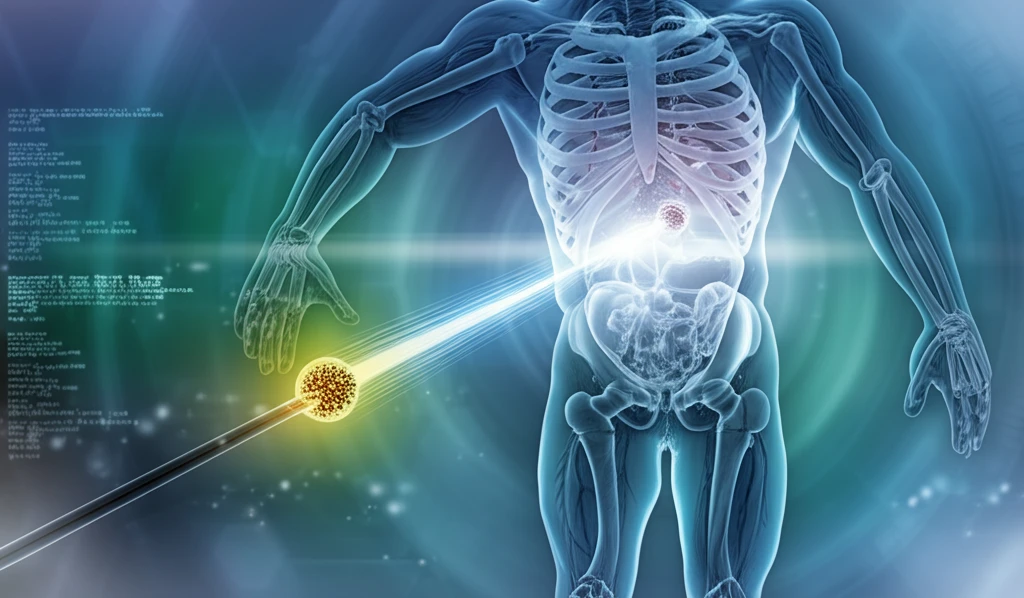
Precision Matters: Ensuring Accuracy in Low-Dose Brachytherapy

"A Deep Dive into the Re-evaluation of Correction Factors for the GROVEX System"
In the realm of medical physics, precision is not merely a goal; it's a necessity. This is especially true in the field of brachytherapy, a form of radiation therapy where radioactive sources are placed inside the body to target cancerous tumors. The accuracy of these treatments hinges on precise measurements and meticulous calculations, ensuring that the radiation dose delivered is exactly what's intended.
Low-dose-rate (LDR) brachytherapy, used for treating cancers like prostate and breast cancer, requires even greater attention to detail. Minute errors in measurement can accumulate, leading to suboptimal treatment or, worse, unintended harm to healthy tissue. Recognizing this, scientists at the Physikalisch-Technische Bundesanstalt (PTB) in Germany have undertaken a critical re-evaluation of the correction factors for the GROVEX (GROssVolumige EXtrapolationskammer) system, a primary standard for LDR brachytherapy dosimetry.
This re-evaluation, detailed in a recent publication, isn't just an academic exercise. It's a practical step towards improving the safety and efficacy of brachytherapy treatments worldwide. By refining the correction factors, the researchers are helping to ensure that medical professionals can deliver the most accurate and effective radiation doses possible.
Measuring Brachytherapy's Reach and Risks
A European survey of facilities and resources provides insights into patterns of care for brachytherapy across the continent, offering statistics on how the technique is delivered and supported regionally. In the prostate setting, an analysis of the time course and accumulated risk of severe urinary adverse events reports numbers needed to harm of 53, 26, 12, and 8 persons, respectively, quantifying how risk accrues with time. A clinical study of applicator insertion used statistical software to collect process time and the incidence of acute side effects during the brachytherapy procedure, underscoring that procedural factors themselves contribute to patient impact. Korean statistics published in the Yonsei Medical Journal also show that the total number of patients treated by HDR brachytherapy has steadily increased, with considerable differences reported between HDR and LDR practice.
Seeds, Applicators, and the Push for Standardization
Low-dose-rate brachytherapy is typically performed as an outpatient procedure in which a physician uses thin needles to implant small radioactive “seeds” within and around the tumor, with as many as 100 or more seeds placed depending on the target. Intracavitary brachytherapy offers a different delivery route, using a specialized applicator inserted into the vagina to deliver radiation directly to the cervix while reducing exposure to healthy tissues. Accurate dosing depends on rigorous dosimetry, and an international code of practice describes primary standards based on graphite cavity ionization chambers, including the BARC method built on the Burlin general cavity theory. Because individualized treatment planning is complex, research indicates that standardizing prostate brachytherapy treatment plans is feasible, with the volume stratified in 10-cc intervals so that most cases appropriate for brachytherapy might be treated with three standard plans.
From Radium to the Golden Age
The origin of brachytherapy is directly related to the discovery of radioactivity by Becquerel in 1896, which led Marie and Pierre Curie to discover radium in 1898 and gave physicians their first practical radioactive sources. Brachytherapy itself is a form of radiation therapy in which a sealed radiation source is placed inside or next to the area requiring treatment, a name derived from the Greek word “brachys,” meaning short. In prostate cancer, a “golden age” of brachytherapy was later documented in historical accounts, a period remembered both for the technique's widespread adoption and for the cautionary lessons drawn from its complications. These milestones trace the arc of brachytherapy from a foundational scientific discovery into a precisely administered clinical tool.
Decoding the GROVEX Re-evaluation
The GROVEX system, in essence, is a highly specialized ionization chamber designed to measure the reference air kerma rate (RAKR) of brachytherapy sources. RAKR is a fundamental quantity that characterizes the strength of a radioactive source. The GROVEX does this by measuring the ionization produced in air by the radiation emitted from the source. However, the raw measurements aren't directly usable; they need to be corrected to account for various factors that can influence the results. These correction factors include:
- Scatter: Radiation bouncing off surfaces like the source holder or collimator.
- Attenuation: Radiation being absorbed by air, filters, or the chamber's components.
- Divergence: The spreading of the radiation beam as it travels from the source.
Advances in Image Guidance and Monotherapy
A recent review provides an overview of state-of-the-art gynecologic brachytherapy, focusing on recent advances and their implications for women with cervical cancer, with image-guided brachytherapy emerging as a central theme. Original research on image-guided interstitial brachytherapy for recurrent disease examines survival after reirradiation, and related work analyzes dose-volume histogram parameters and late side effects in magnetic resonance image-guided adaptive cervical cancer brachytherapy. For localized prostate cancer, a study of high-dose-rate brachytherapy used as monotherapy reported late Grade 2 gastrointestinal toxicity of just 0.4%, with no Grade 3 or greater late adverse events. Ongoing coverage at Medical Xpress highlights remaining clinical questions, such as the best way to treat recurring prostate cancer.
Documentation, Relapses, and the Other Side of the Story
Clinicians stress that brachytherapy succeeds or fails on process discipline: proper documentation, including radiotherapy prescriptions, written directives for physicists, and implant checklists, is described as critical to ensuring all parameters are met and safety is maintained. Outcomes data also record failures: a critical mid-term evaluation of 250 prostate I-125 brachytherapy cases found a biochemical failure (by Phoenix criteria) in 10 patients (6.6%), with prostate biopsy confirming cancer in 6 who underwent radical prostatectomy (4) or external radiotherapy (2), and one patient developing systemic progression with secondary bone lesions. That same study, reported separately in the literature, nonetheless concluded that brachytherapy in low-grade risk prostate cancer represents a good alternative to radical prostatectomy, with excellent functional and oncologic results. Patient accounts add a personal layer, with one Mayo Clinic Connect contributor reporting zero problems with urinary urgency and no issues with erectile dysfunction.
Brachytherapy vs. Surgery, EBRT, and IMRT
Comparative analyses suggest brachytherapy offers similar cancer control to alternatives for many early-stage cancers while delivering better functional and cosmetic results. An indirect comparison of observational studies has suggested that overall survival rates with LDR brachytherapy are similar to those with alternatives including radical prostatectomy and external beam radiation therapy. In gynaecological cancer, comparisons of IMRT and brachytherapy note that the role of radiotherapy and brachytherapy in the management of locally advanced cervical and endometrial cancer is well established. Brachytherapy's central advantage is often framed as precision: it is designed to target cancer with high dose intensity while minimizing damage to healthy tissues and reducing side effects compared with external radiation therapy.
The Impact on Patient Care
While the technical details of the GROVEX re-evaluation might seem arcane, the implications for patient care are very tangible. The researchers found that the updated correction factors led to a decrease of approximately 0.9% in the measured reference air kerma rate for a representative seed of type Bebig I25.S16C. This might sound like a small change, but in the world of radiation therapy, even small adjustments can have a significant impact on treatment outcomes. By incorporating these refined correction factors, medical physicists can more accurately calculate the radiation dose delivered to the tumor, optimizing the treatment plan and minimizing the risk of side effects. This meticulous approach underscores the commitment to precision and patient safety that drives progress in medical physics.
Expert Views: From DNA Damage to Electronic Sources
Memorial Sloan Kettering explains that brachytherapy works by implanting radioactive material sealed inside a seed, pellet, wire, or capsule using a needle or catheter, with the radiation damaging the DNA of nearby cancer cells. Patient-facing experts note that some analyses show HDR brachytherapy monotherapy achieving competitive results with triple-modality therapy, an attractive option for patients still deciding on androgen deprivation therapy. In China, a formal expert consensus was developed for computed tomography-guided iodine-125 radioactive seed permanent interstitial brachytherapy, with the evidence analyzed and the opinions and suggestions of experts synthesized into guidance. The American Brachytherapy Society has also published its first consensus statement on electronic brachytherapy, which uses electrically generated X-rays instead of radioactive isotopes, a development clinicians should understand.
A Growing, Technology-Driven Field
Market analysis projects the brachytherapy market will grow at a compound annual growth rate of 7.3%, reaching USD 3.3 billion by 2035, while the devices segment is described as a growing part of the medical industry driven by advancements in cancer treatment technologies. Key trends shaping the catheter market include the shift toward image-guided brachytherapy, increasing adoption of high-dose-rate brachytherapy, and development of advanced catheter designs. Researchers credit the dramatic advances of the last decade largely to improvements in applicators, imaging, treatment planning, and use of clinical trials, and note that current research continues to change how the modality is delivered. Together, these projections and technical developments point to continued growth for brachytherapy as a treatment option.
Building Programs and Facing Systemic Barriers
Real-world studies, such as one conducted at the Bahawalpur Institute of Nuclear Medicine and Oncology in Pakistan, evaluate how the geometry of brachytherapy applicators affects delivered dose in intracavitary cervical cancer treatment, underscoring the technical precision required. Establishing new brachytherapy programs is a substantial undertaking: an Omani cancer center report stratified the challenges of implementing an interstitial needle brachytherapy program into equipment, expertise, quality, patient compliance, and importation of radioactive sources. Predictive analytics is being explored as a way to optimize clinical outcomes in this highly targeted form of internal radiation therapy for localized cervical, prostate, and breast cancers. Broader access questions remain as well, since a systematic review has evaluated the feasibility of modern external beam radiotherapy as an alternative approach to brachytherapy in endometrial cancer treatment.
Real Clinics, Real Patients, and Smarter Planning
On the technology side, vendors report real-time precision in HDR prostate brachytherapy, with Siemens Healthineers describing how Advanced Oncology Solutions helped CHRISTUS Health accelerate the adoption of tech-enabled radiation-oncology services. In cervical cancer, a preliminary study suggests that tumor perfusion micro-topography features revealed by DCE-MRI influence primary dose response, with the tumor peripheral region more relevant than the central region, which could potentially affect treatment plan design. Planning software such as Elekta's Oncentra Brachy enables real 3D contouring by combining arbitrary-plane views with tools such as the pearl tool. For regional health networks, AI-assisted brachytherapy planning offers competitive advantages, including faster treatment cycles and improved patient satisfaction, which in turn enhance overall care quality.