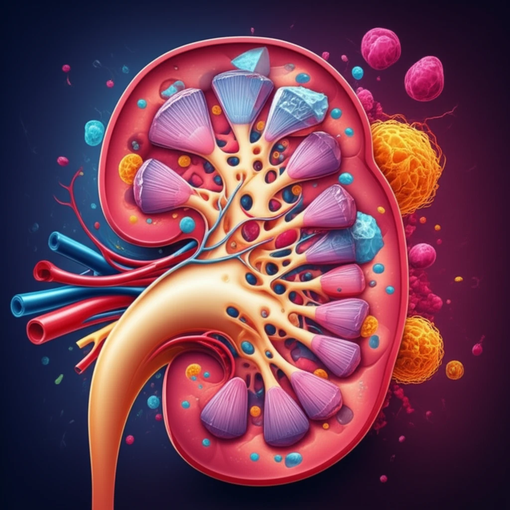
Surreal illustration of kidney stone formation with rapidly multiplying cells.

Kidney Stones: Is Your Body Triggering Its Own Crystal Crisis?

"New research reveals how urothelial proliferation in the kidneys could be a key factor in the development of renal crystal deposits, offering potential insights for prevention."


Kidney stones, a common ailment affecting a significant portion of the population, are often attributed to factors like diet and hydration. However, emerging research suggests a more complex picture, highlighting the role of processes within the kidney itself.

A recent study delved into the early events of kidney stone formation, specifically focusing on how crystals bind to the inner lining of the kidneys, known as the urothelium. The findings suggest that urothelial proliferation, or the rapid growth of these cells, may be a critical trigger in the development of renal crystal deposits.

This article explores the surprising link between urothelial proliferation and kidney stone formation, drawing on the study's insights to shed light on potential preventative strategies and the future of kidney stone research.

The Urothelium-Crystal Connection: How Cell Growth Can Lead to Stone Formation

Surreal illustration of kidney stone formation with rapidly multiplying cells.

The study, conducted on a murine model, investigated the effects of vitamin D supplements, along with water containing hydroxyl-L-proline, ammonium chloride, and calcium chloride, on kidney crystal formation. Researchers compared a group receiving Fibroblast Growth Factor 7 (FGF7), a mitogen that stimulates urothelial cell growth, with a control group.

The results revealed that calcium oxalate monohydrate (COM) crystals were detected in the urine as early as day 2, with enhanced deposits in the FGF7 group by day 14. This acceleration suggests a direct link between urothelial proliferation and crystal deposition.

  • Urothelial Proliferation: The FGF7 group exhibited significant urothelial cell proliferation, indicating rapid cell growth in the kidney lining.
  • Uroplakin III Downregulation: A decrease in uroplakin III, a protein found in urothelial cells, was observed in the FGF7 group.
  • CD44 Expression: The FGF7 group showed de novo expression of osteopontin receptor CD44, which plays a role in cell adhesion and crystal retention.
These changes suggest that urothelial proliferation leads to alterations in cell characteristics, making it easier for crystals to adhere and accumulate. Interestingly, when the mice were switched back to a normal diet on day 21, a spontaneous crystal clearance was observed, underscoring the influence of external factors on crystal formation.

What This Means for You: Preventing Crystal Buildup and Future Research

The study's findings highlight the importance of maintaining a healthy kidney environment to prevent excessive urothelial proliferation. While the research was conducted on animal models, the implications for human health are significant.

Future research should focus on identifying specific factors that trigger urothelial proliferation in the kidneys, as well as exploring potential therapeutic interventions to regulate cell growth and prevent crystal adhesion. Understanding these mechanisms could lead to more targeted and effective strategies for preventing kidney stone formation.

By understanding the interplay between urothelial proliferation and crystal formation, we can move closer to personalized strategies for preventing kidney stones and promoting long-term renal health.

About this Article -

This article was crafted using a human-AI hybrid and collaborative approach. AI assisted our team with initial drafting, research insights, identifying key questions, and image generation. Our human editors guided topic selection, defined the angle, structured the content, ensured factual accuracy and relevance, refined the tone, and conducted thorough editing to deliver helpful, high-quality information.See our About page for more information.

This article is based on research published under:

DOI-LINK: 10.1038/s41598-018-34734-8, Alternate LINK

Title: Urothelium Proliferation Is A Trigger For Renal Crystal Deposits In A Murine Lithogenesis Model

Subject: Multidisciplinary

Journal: Scientific Reports

Publisher: Springer Science and Business Media LLC

Authors: Héloïse Bilbault, Joëlle Perez, Léa Huguet, Sophie Vandermeersch, Sandrine Placier, Nahid Tabibzadeh, Vincent Frochot, Emmanuel Letavernier, Dominique Bazin, Michel Daudon, Jean-Philippe Haymann

Published: 2018-11-05

Everything You Need To Know

1

What is the main focus of the new research regarding kidney stones?

Kidney stones, a common ailment, are often linked to diet and hydration. However, research points to the inner workings of the kidneys. The findings suggest that **urothelial proliferation**, or rapid cell growth in the kidney lining, may be a crucial factor in the formation of these deposits. The article suggests that the kidneys might play a more direct role than previously understood, highlighting internal processes as potential targets for prevention.

2

How does urothelial proliferation contribute to kidney stone formation?

The research indicates that **urothelial proliferation** seems to be a key factor in kidney stone development. This is because the rapid cell growth in the kidney lining alters the environment, making it easier for crystals to stick and accumulate. This was observed in a study where a group of mice received **Fibroblast Growth Factor 7 (FGF7)**, a mitogen that stimulates **urothelial cell growth**, leading to increased crystal deposits. The implications are that if the rapid cell growth can be controlled, the chances of crystal buildup might be reduced.

3

What specific cellular changes occur during urothelial proliferation that promote crystal formation?

The study focused on the **urothelium**, the inner lining of the kidneys, and how it interacts with crystals. The study showed that when the **urothelial proliferation** occurs, there are changes in cell characteristics. In the **FGF7** group, the **urothelial cells** exhibited a decrease in **uroplakin III**, a protein found in the **urothelial cells**. The FGF7 group also showed de novo expression of **CD44**, which aids in crystal retention. These alterations make it easier for crystals, like **calcium oxalate monohydrate (COM)**, to attach and grow. The implications are that strategies targeting these cellular changes could prevent crystal buildup.

4

What are the broader implications of this research for preventing kidney stones?

The implications of this research are significant for understanding and preventing kidney stones. The study highlighted that **urothelial proliferation** and the resulting cellular changes are linked to the buildup of crystals. The study found that when the mice were switched back to a normal diet, the **COM** crystal deposits were spontaneously cleared. This underscores the importance of a healthy kidney environment and the potential for dietary or other interventions to influence crystal formation and dissolution. The study suggests that maintaining a healthy kidney environment is crucial to prevent excessive **urothelial proliferation** and crystal buildup.

5

What was the experimental setup in this research?

The research underscores the importance of the **urothelium** and internal processes in kidney stone formation. The study introduced **vitamin D supplements**, along with water containing **hydroxyl-L-proline, ammonium chloride, and calcium chloride**, which led to **COM** crystal formation. The study then administered **Fibroblast Growth Factor 7 (FGF7)**, which promotes **urothelial proliferation** and crystal deposition. The mice that switched back to a normal diet showed crystal clearance. Researchers are also studying how crystals bind to the **urothelium** and the role of the protein **uroplakin III** and **CD44** expression in crystal retention. These findings suggest various research paths.

Newsletter Subscribe

Subscribe to get the latest articles and insights directly in your inbox.