
Keloid or Not? How to Spot Cutaneous Leiomyomas Mimicking Keloids

"Uncover the key differences between keloids and cutaneous leiomyomas and ensure accurate diagnosis."
Cutaneous leiomyomas are rare, benign tumors arising from the smooth muscle of the skin. While harmless, they can sometimes be mistaken for other skin conditions, most notably keloids. Keloids, on the other hand, are abnormal proliferations of scar tissue that develop after skin injuries, surgery, or trauma.
Distinguishing between these two conditions is crucial for appropriate management. Keloids typically grow beyond the original boundaries of the wound, while hypertrophic scars remain confined. But what happens when a benign tumor starts acting like a scar?
This article delves into a fascinating case study that highlights the diagnostic challenges posed by cutaneous leiomyomas mimicking keloids. We'll explore the clinical features, diagnostic approaches, and why early recognition is essential, potentially even life-saving.
What Keloid Research Measures
A December 8, 2023, real-world retrospective cohort study set out to characterize patients with keloids in a large US-based cohort, comparing them with matched dermatology and general cohorts. It used structured and unstructured secondary data to examine keloid severity and progression, including differences by sociodemographic factors. The supplied study descriptions do not provide prevalence or other numerical estimates, so they do not support a specific population-level statistic.
Three Types of Cutaneous Leiomyoma
A 2016 report on cutaneous leiomyomas mimicking keloids describes three types according to tumor origin: piloleiomyomas, angioleiomyomas, and genital leiomyomas. This classification provides a basic framework for describing these smooth-muscle tumors. The supplied source does not specify when the classification was first established or detail earlier milestones.
The Case of the Misdiagnosed Shoulder Keloid: A Diagnostic Journey
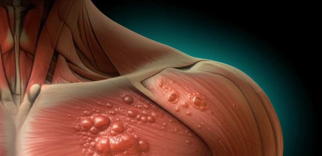
Imagine a 23-year-old woman seeking treatment for what appears to be keloids on her left shoulder. She’s had these raised skin lesions since childhood, experiencing mild itching and discomfort. A general practitioner initially diagnosed keloids, a reasonable assumption given the appearance and location.
- Clinical Appearance: Raised skin lesions, potentially reddish-brown in color, resembling keloids.
- Patient History: Absence of typical keloid triggers like significant trauma or surgery.
- Location: While keloids often appear on shoulders, other locations can raise suspicion.
Reviews Emphasize Diagnosis and Evidence Gaps
An August 2025 review, “Keloids revisited: Current concepts in treatment and differential diagnosis,” aims to summarize current treatment concepts and differential diagnosis to help clinicians and researchers improve diagnostic accuracy and select personalized treatments. A September 2025 review reports that its literature search found no keloid-treatment meta-analysis, one randomized clinical trial, and two systematic reviews. A February 2026 case report notes that biopsy may be needed to investigate other causes when standard treatment, such as intralesional steroid injections, does not work for a presumed hypertrophic scar or keloid.
A Keloid-Like Lesion May Have Another Cause
Cutaneous leiomyomas may be mistaken for keloids, and the 2016 report notes their clinical relevance because they may signal hereditary leiomyomatosis and renal cell cancer. One case description reports red-brown, firm nodules with epidermal thinning that clinically resembled keloids; another patient with pilar leiomyoma was reportedly treated with cryotherapy and intralesional steroid injections for presumed eruptive keloids before histopathology established the diagnosis. A separate report describes cutaneous leiomyosarcoma as a rare tumor type representing 1%–3% of all sarcomas and stresses the importance of early diagnosis for successful management and curative intent.
Why Accurate Diagnosis Matters: Beyond the Cosmetic
While cutaneous leiomyomas are benign, recognizing them is clinically relevant. These tumors can be associated with hereditary leiomyomatosis and renal cell carcinoma (HLRCC), also known as Reed’s syndrome. This genetic condition increases the risk of developing uterine fibroids, skin leiomyomas, and a specific type of kidney cancer.
Misleading Features Can Delay Recognition
A 2018 case report describes a delayed diagnosis of dermal leiomyosarcoma that mimicked a keloid scar in a patient who had previously had a leiomyoma excised from the same site. The report identifies misleading lesion features, neglected lumps, and inaccurate diagnosis as possible reasons for delay. It also recounts a pilar leiomyoma that was treated as an eruptive keloid with cryotherapy and intralesional steroid injections before histopathologic verification.
Research Needs and Market Projection
A 2024 review says keloid pathogenesis is not fully elucidated and identifies the absence of standardized guidelines or treatment protocols endorsed by academic governing bodies as a continuing challenge. It examines molecular and cellular mechanisms underlying keloid formation, reflecting an ongoing effort to clarify the condition. Separately, a market report dated September 15, 2026, projects the keloid-treatment market will reach USD 1.9 billion by 2035, with a 0.86% compound annual growth rate from 2025 to 2035.
Quality of Life and Uneven Evidence
A 2024 article describes keloids as prevalent but understudied and commonly affecting people of color; it notes that keloids can negatively affect many aspects of patients’ quality of life, while the impact has not been comprehensively characterized. A separate review says systemic diseases, general health conditions, genetic disorders, lifestyle, and the natural environment may contribute to keloid severity and risk. A 2026 systematic-review abstract notes that keloid scars disproportionately affect Black and Asian populations, but says evidence on psychosocial impacts across diverse patient populations remains limited and measurement approaches vary.
A Painful Shoulder Lesion Misdiagnosed as Keloid
A case report describes a 26-year-old woman with Fitzpatrick skin type 4/5 who visited a dermatology outpatient department for treatment of a painful lesion on her left shoulder. The cutaneous leiomyoma had clinical similarities to a keloid and had previously been misdiagnosed and managed as one. The case illustrates how resemblance in appearance can affect a patient’s treatment course.
In the presented case, the patient was fortunate. Screening for uterine leiomyoma and renal cell carcinoma came back negative. However, early diagnosis of cutaneous leiomyomas allows for timely investigations and monitoring for potential underlying genetic conditions.
So, next time you encounter a suspected keloid, remember that it could be something else. A thorough examination, a detailed patient history, and, when necessary, a skin biopsy can make all the difference. Early recognition and accurate diagnosis aren't just about aesthetics; they can be life-saving.