Intraoperative Ultrasound: The Surgeon's Sixth Sense in Congenital Hyperinsulinism
"Discover how real-time ultrasound is revolutionizing tissue-sparing surgery for children with congenital hyperinsulinism, minimizing complications and maximizing cure rates."
Congenital hyperinsulinism (CHI) is a rare genetic disorder where the pancreas secretes too much insulin, leading to persistent hypoglycemia, especially in infants. This condition can cause seizures, brain damage, and developmental delays if not promptly and effectively treated. The challenge lies in identifying and removing only the affected part of the pancreas, preserving as much healthy tissue as possible.
Traditionally, surgeons relied on preoperative imaging and intraoperative palpation (feeling with their hands) to locate problematic areas within the pancreas. However, these methods can be imprecise, particularly when the lesions are small or deep within the tissue. Removing too much of the pancreas can lead to diabetes later in life, making precise surgery critical.
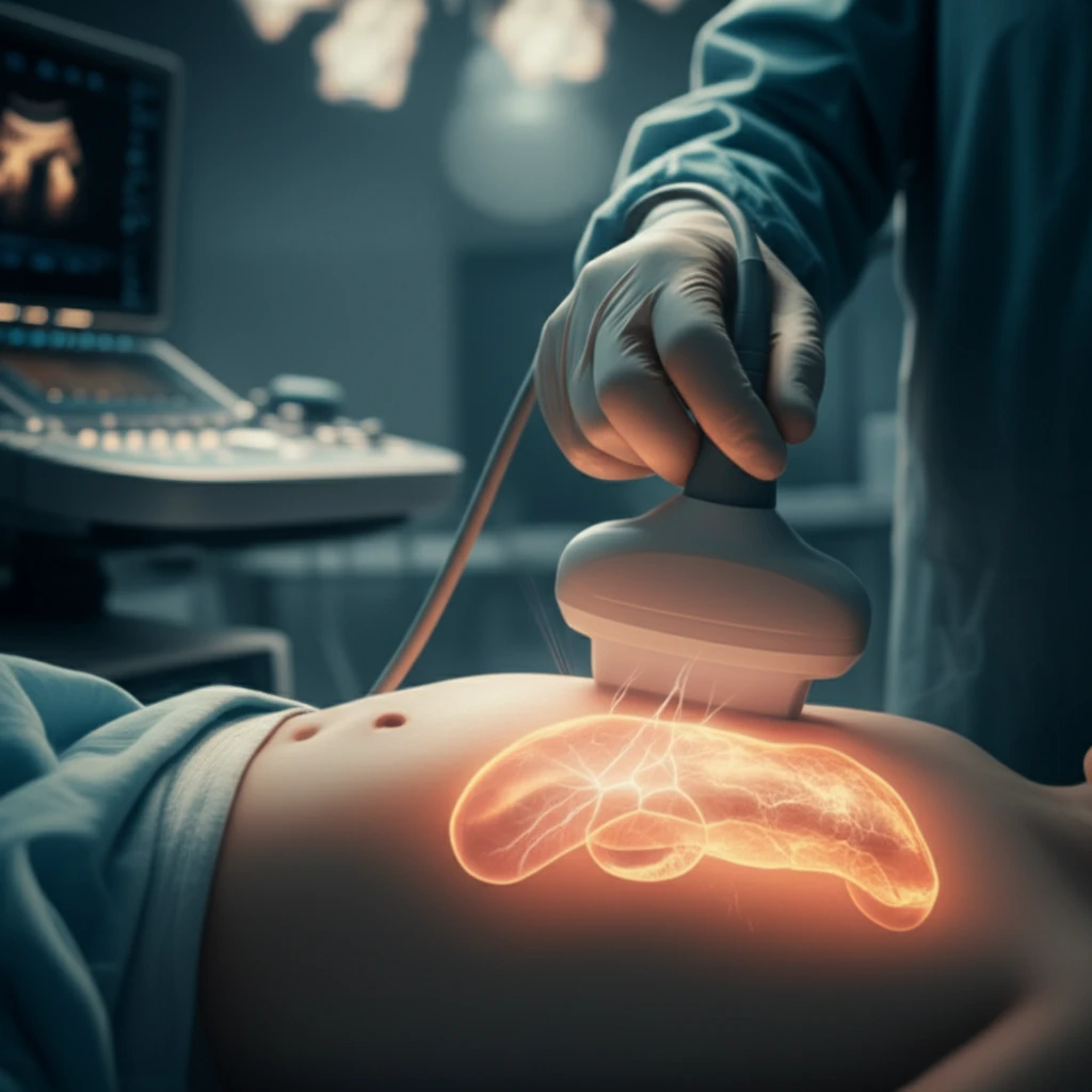
Enter intraoperative ultrasound (IOUS), a real-time imaging technique that allows surgeons to visualize the pancreas during the operation. Think of it as a surgeon's sixth sense, providing a live view of what lies beneath the surface. This technology promises to enhance the precision of tissue-sparing surgery, improving outcomes for children with focal CHI.
How Does Intraoperative Ultrasound Change the Game?
A recent study published in "Frontiers in Endocrinology" investigated the use of IOUS in guiding surgical resections for children with focal CHI. Researchers at Odense University Hospital in Denmark conducted a retrospective analysis of 24 patients with focal CHI who underwent surgery between 2010 and 2017. All patients had confirmed focal lesions via genetic testing and 18F-DOPA-PET/CT scans.
- Enhanced Localization: IOUS successfully located the focal lesion in 16 out of 20 patients (80% sensitivity).
- Improved Specificity: IOUS correctly identified the absence of focal lesions in all 11 patients with diffuse CHI (100% specificity).
- Tissue-Sparing Success: Guided by IOUS, surgeons performed tissue-sparing resections in 67% of patients, removing only the affected tissue.
- Ectopic Lesion Detection: IOUS aided in locating an ectopic lesion (a lesion outside the normal location) in the duodenal wall, a rare occurrence that can be challenging to identify.
The Future of CHI Surgery
This study adds to the growing body of evidence supporting the use of IOUS as a valuable tool in CHI surgery. By providing real-time visualization, IOUS enables surgeons to perform more precise and less invasive resections, leading to better outcomes for children with this challenging condition. The combination of genetics, advanced imaging, and innovative surgical techniques offers new hope for a future where CHI can be effectively managed with minimal long-term consequences.