DNA strand intertwined with an iron-sulfur cluster.

DNA Primase's Redox Role: New Insights into Enzyme Activity

"A fresh look at the structure of the iron-sulfur cluster domain in DNA primase challenges previous assumptions about its redox function and suggests alternative explanations for enzyme activity."


The process of DNA replication relies on a complex interplay of enzymes, each with a specific role. Among these, DNA primase is responsible for synthesizing short RNA primers that initiate DNA synthesis. Recent research by O'Brien et al. (1) proposed that the iron-sulfur cluster within primase has a redox function, influencing its activity.

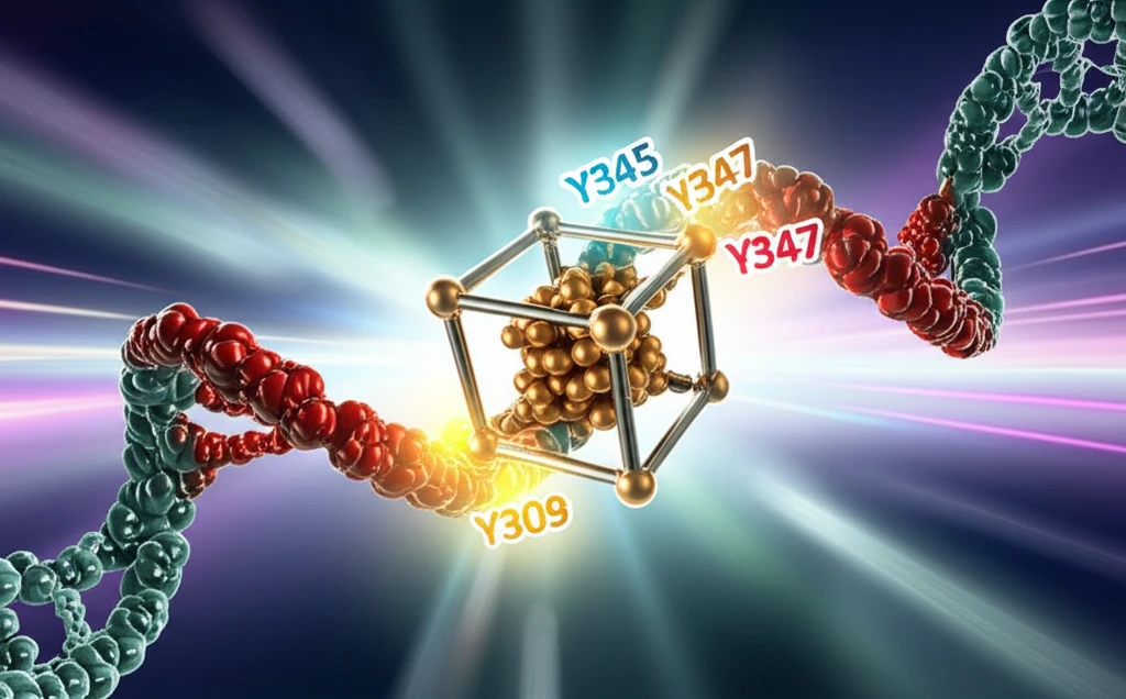
The study suggested that the redox state of this cluster acts as a switch, modulating DNA binding and primer termination. The proposed mechanism involved electron transfer through the DNA helix, facilitated by specific tyrosine residues in primase. However, this model hinges on a particular structure of the primase domain, which has been called into question.

This article delves into a structural re-evaluation of the primase domain, revealing a potentially misfolded structure in the original study. We will explore how the corrected structure alters the understanding of the tyrosine-mediated electron transfer pathway and offers alternative explanations for primase's function in DNA replication.

The Misfolded Structure and Its Implications

DNA strand intertwined with an iron-sulfur cluster.

O'Brien et al.'s (1) hypothesis of the Fe-S cluster's redox activity hinges on a specific structure of the primase domain (p58C). This structure, however, contains a local misfolding—amino acids 318 to 360 adopt a beta-hairpin conformation instead of the alpha-helical structure observed in other structures of yeast and human p58C (4, 5). This discrepancy is likely due to a single-point mutation (I271S) present in the p58C construct used in the original study (3).

The correct alpha-helical structure dramatically changes the positions of key tyrosine residues (345 and 347) thought to be involved in electron transfer. The distance between these residues and another tyrosine (309) becomes significantly larger (Fig. 1B). This raises doubts about the feasibility of electron transfer between these residues on a physiologically relevant timescale.

Here's a breakdown of the structural differences and their functional implications:
  • Incorrect Folding: The beta-hairpin conformation alters the spatial arrangement of critical amino acids.
  • Increased Distance: The distance between tyrosine residues increases, potentially hindering electron transfer.
  • Non-conserved Residue: Tyrosine 309, a key component of the proposed electron wire, is not conserved across all eukaryotic primases.
A recent structural analysis of human p58C bound to an RNA primer/DNA template further supports this revised understanding (6). This structure reveals that tyrosines 345 and 347 are located at the interface with the RNA/DNA helix, interacting with the triphosphate group at the 5' end of the RNA primer and the DNA template. Therefore, any changes in primase activity observed in the Y345 mutant may be due to weakened RNA/DNA binding rather than impaired charge transfer.

Reassessing the Redox Role of Primase

Given the structural concerns and the alternative explanation for the mutagenesis data presented by O'Brien et al., it's important to reconsider the proposed redox role of the Fe-S cluster in primase. The evidence supporting this role appears less definitive when viewed through the lens of the corrected protein structure.

While the idea of a redox switch in primase is intriguing, further experimentation is needed to confirm this mechanism. Future studies should focus on validating the electron transfer pathway and directly measuring the redox potential of the Fe-S cluster in the context of the correctly folded primase structure.

Understanding the precise function of the iron-sulfur cluster in primase remains an open question. By considering the accurate protein structure and exploring alternative explanations, researchers can continue to unravel the complexities of DNA replication and the roles of key enzymes like primase.

About this Article -

This article was crafted using a human-AI hybrid and collaborative approach. AI assisted our team with initial drafting, research insights, identifying key questions, and image generation. Our human editors guided topic selection, defined the angle, structured the content, ensured factual accuracy and relevance, refined the tone, and conducted thorough editing to deliver helpful, high-quality information.See our About page for more information.

This article is based on research published under:

DOI-LINK: 10.1126/science.aan2954, Alternate LINK

Title: Comment On “The [4Fe4S] Cluster Of Human Dna Primase Functions As A Redox Switch Using Dna Charge Transport”

Subject: Multidisciplinary

Journal: Science

Publisher: American Association for the Advancement of Science (AAAS)

Authors: Luca Pellegrini

Published: 2017-07-21

Everything You Need To Know

1

What is the role of DNA primase, and how does it relate to the iron-sulfur cluster?

DNA primase synthesizes short RNA primers, acting as the initiator in DNA replication. The iron-sulfur cluster within DNA primase was initially proposed to have a redox function, influencing its activity by modulating DNA binding and primer termination. The correct protein folding is essential for understanding the precise role of the Fe-S cluster and tyrosine residues in the electron transfer process.

2

Why is the structure of the iron-sulfur cluster domain in DNA primase important?

The structure of the iron-sulfur cluster domain in DNA primase is vital because it dictates the spatial arrangement of amino acids, especially the tyrosine residues. Incorrect folding, like the beta-hairpin conformation, can alter the distances between these residues, potentially disrupting electron transfer. The alpha-helical structure, supported by recent structural analyses, suggests an alternative role for these tyrosines, focusing on RNA/DNA binding.

3

How does a misfolded structure impact the understanding of DNA primase's function?

A misfolded structure, specifically a beta-hairpin conformation in the primase domain (p58C) due to a mutation (I271S), challenges the original hypothesis. This misfolding affects the positions of key tyrosine residues (345 and 347), increasing the distance between them and tyrosine 309. This makes the electron transfer process less feasible. The alpha-helical conformation in other primase structures supports this reevaluation, which suggests that the tyrosine residues might be involved in RNA/DNA binding instead of electron transfer.

4

What role were tyrosine residues thought to play in DNA primase, and how has this been reevaluated?

Tyrosine residues, especially tyrosines 345, 347 and 309, were initially hypothesized to be involved in electron transfer within DNA primase. However, their actual role has been reevaluated due to structural analysis. In the correct alpha-helical structure, these tyrosines are located at the interface with the RNA/DNA helix, interacting with the primer and template. This suggests that their function may be related to RNA/DNA binding rather than mediating electron transfer.

5

What are the implications of this revised understanding of DNA primase's structure and function?

The implications of the revised understanding of DNA primase's structure and function are significant. The initial proposal suggested a redox role for the Fe-S cluster; however, with the evidence pointing to misfolded structures, that theory has been reevaluated. The new structural data, particularly the alpha-helical conformation, alters the understanding of the tyrosine-mediated electron transfer pathway. This shift impacts how we interpret experiments and understand DNA primase's overall function during DNA replication.

Newsletter Subscribe

Subscribe to get the latest articles and insights directly in your inbox.