Hyperdense Middle Cerebral Artery Sign (HMCAS) on a brain scan.

Decoding the Hyperdense Middle Cerebral Artery Sign: What It Means After Trauma

"A closer look at HMCAS, its implications for stroke patients, and the latest in treatment strategies."


Imagine a scenario: a patient arrives at the emergency department following a high-speed motor vehicle accident. Physical examination reveals no immediate signs of head trauma, yet something isn't right. The patient exhibits a Glasgow Coma Scale score of 6, indicating impaired consciousness and responsiveness. Computed tomography (CT) scans reveal a hyperdense middle cerebral artery sign (HMCAS) on one side of the brain.

This scenario, drawn from a real case study, highlights the critical importance of recognizing and understanding HMCAS, particularly in the context of trauma. HMCAS is a radiological finding that suggests a blood clot or thrombus within the middle cerebral artery, potentially leading to stroke and severe neurological deficits.

In this article, we'll delve into the intricacies of HMCAS, exploring its causes, diagnostic methods, and the latest treatment strategies. We'll also discuss how early identification and intervention can significantly improve outcomes for patients who present with this ominous sign.

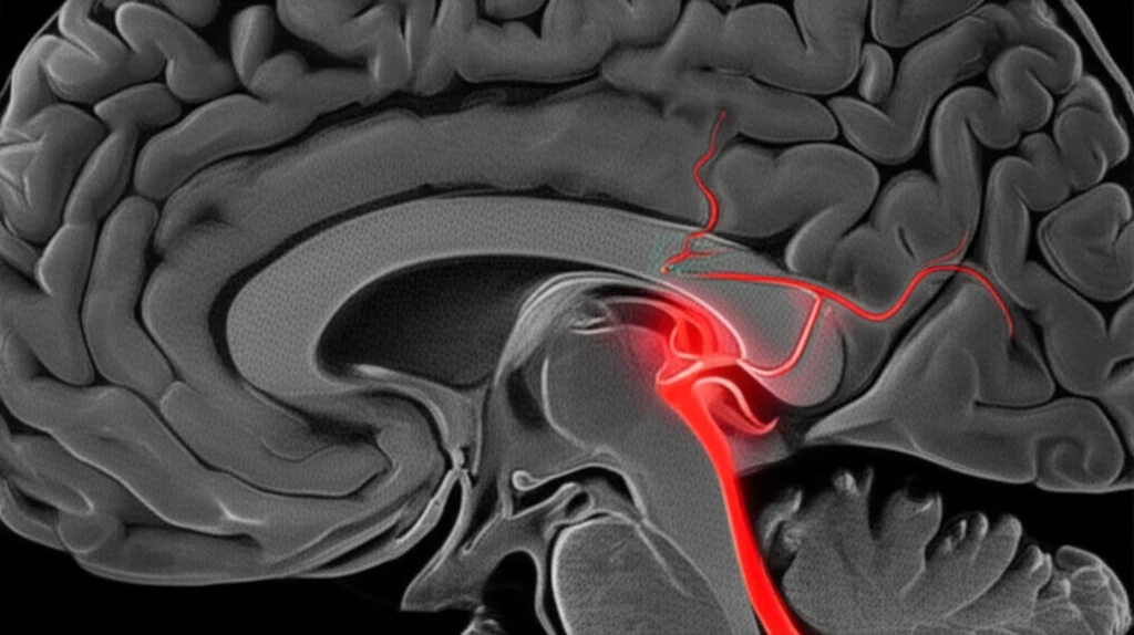
What is the Hyperdense Middle Cerebral Artery Sign (HMCAS)?

Hyperdense Middle Cerebral Artery Sign (HMCAS) on a brain scan.

The Hyperdense Middle Cerebral Artery Sign (HMCAS) is a radiological finding observed on computed tomography (CT) scans of the brain. It appears as an unusually dense or bright area within the middle cerebral artery (MCA), one of the major blood vessels supplying blood to the brain.

This hyperdensity indicates the presence of a blood clot or thrombus within the artery. The increased density is due to the high concentration of hemoglobin in the clotted blood, which attenuates X-rays more strongly than normal blood or surrounding brain tissue. The occurrence of HMCAS is typically associated with acute ischemic stroke, where a sudden blockage of blood flow deprives brain tissue of oxygen and nutrients.

  • Early Detection: Recognizing HMCAS on initial CT scans is crucial for prompt diagnosis and intervention.
  • Stroke Indicator: It strongly suggests a blockage in the MCA, warranting further investigation.
  • Treatment Decisions: HMCAS can influence treatment strategies, such as thrombolysis or thrombectomy.
When HMCAS is identified, it triggers a series of actions to confirm the diagnosis and determine the best course of action. This typically involves additional imaging, such as magnetic resonance imaging (MRI) or angiography, to visualize the blood vessels and assess the extent of the blockage. Neurological assessments are also performed to evaluate the patient's neurological function and identify any deficits.

The Future of HMCAS Research and Treatment

Ongoing research continues to refine our understanding of HMCAS and its implications for stroke management. Studies are investigating new imaging techniques, such as advanced CT perfusion and MRI sequences, to improve the detection and characterization of HMCAS. Clinical trials are also evaluating novel therapies, including next-generation thrombolytic agents and mechanical thrombectomy devices, to enhance the effectiveness of stroke treatment. Ultimately, the goal is to develop more personalized and targeted approaches to stroke care, optimizing outcomes for each individual patient.

About this Article -

This article was crafted using a human-AI hybrid and collaborative approach. AI assisted our team with initial drafting, research insights, identifying key questions, and image generation. Our human editors guided topic selection, defined the angle, structured the content, ensured factual accuracy and relevance, refined the tone, and conducted thorough editing to deliver helpful, high-quality information.See our About page for more information.

This article is based on research published under:

DOI-LINK: 10.7556/jaoa.2014.175, Alternate LINK

Title: Hyperdense Middle Cerebral Artery Sign After Trauma

Subject: Complementary and alternative medicine

Journal: Journal of Osteopathic Medicine

Publisher: Walter de Gruyter GmbH

Authors: Robert T. Dahlquist, Ronald F. Sing

Published: 2014-11-01

Everything You Need To Know

1

What exactly is the Hyperdense Middle Cerebral Artery Sign (HMCAS)?

The Hyperdense Middle Cerebral Artery Sign (HMCAS) is a radiological finding, observed on computed tomography (CT) scans. It appears as an area of increased density within the Middle Cerebral Artery (MCA), a major blood vessel supplying the brain. This increased density is due to a blood clot or thrombus, indicating a potential blockage and leading to stroke.

2

Why is identifying the Hyperdense Middle Cerebral Artery Sign (HMCAS) so important?

The Hyperdense Middle Cerebral Artery Sign (HMCAS) is critical because it strongly suggests a blockage in the Middle Cerebral Artery (MCA), which can cause an ischemic stroke. This is significant as it deprives brain tissue of oxygen and nutrients. Early identification of HMCAS allows for prompt intervention, such as thrombolysis or thrombectomy, which can significantly improve patient outcomes. This is especially vital after trauma due to the potential for blood clots forming in the injured patient.

3

What typically causes the Hyperdense Middle Cerebral Artery Sign (HMCAS)?

The causes of the Hyperdense Middle Cerebral Artery Sign (HMCAS) are primarily related to blood clots or thrombi within the Middle Cerebral Artery (MCA). These clots can form due to various factors, including trauma, which can damage blood vessels and increase the risk of clot formation. When a patient experiences a high-speed motor vehicle accident, for example, the trauma can cause the blood vessels to become damaged. The body responds by initiating a clotting cascade, and these clots can then lead to HMCAS.

4

What happens if someone is found to have the Hyperdense Middle Cerebral Artery Sign (HMCAS)?

If someone is found to have the Hyperdense Middle Cerebral Artery Sign (HMCAS), it triggers a series of actions. Additional imaging such as magnetic resonance imaging (MRI) or angiography is used to visualize the blood vessels and assess the extent of the blockage. Neurological assessments are also performed to evaluate the patient's neurological function and identify any deficits. Treatment strategies, such as thrombolysis or thrombectomy, will be considered, depending on the severity and location of the clot.

5

What are the current treatments and future advancements for the Hyperdense Middle Cerebral Artery Sign (HMCAS)?

Current treatments for the Hyperdense Middle Cerebral Artery Sign (HMCAS) involve therapies aimed at removing or dissolving the blood clot, and include: thrombolysis, which uses medications to break down the clot, and thrombectomy, which is a mechanical procedure to remove the clot from the Middle Cerebral Artery (MCA). Future research focuses on new imaging techniques and therapies to personalize stroke care and optimize patient outcomes. Advanced CT perfusion and MRI sequences will help with better detection of HMCAS, and clinical trials evaluate next-generation thrombolytic agents and mechanical thrombectomy devices.

Newsletter Subscribe

Subscribe to get the latest articles and insights directly in your inbox.