
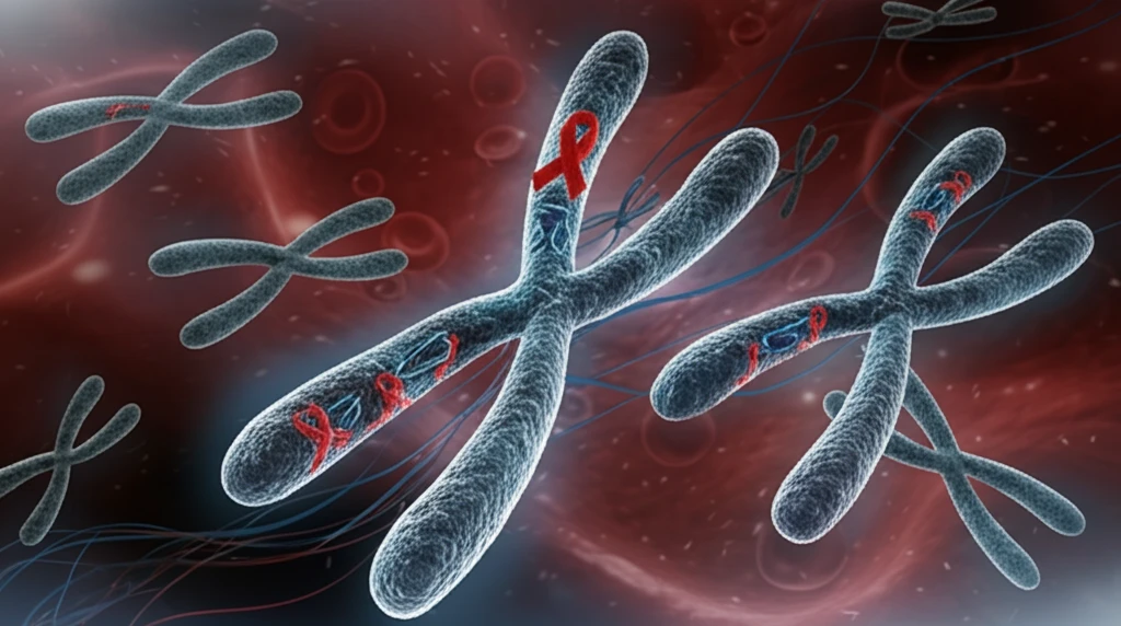
Decoding the Genetic Puzzle: How Chromosome Rearrangements Impact Blood Cancer

"Unraveling the link between t(3;8)(q26.2;q24) translocation and therapy-related myeloid neoplasms for better prognosis."
In the complex world of genetics, chromosomal rearrangements can act as significant drivers of disease, particularly in cancer. Among these, the translocation t(3;8)(q26.2;q24) stands out as a rare yet critical abnormality associated with myeloid neoplasms, a group of cancers affecting the blood and bone marrow. This translocation involves the exchange of genetic material between chromosomes 3 and 8 at specific locations, leading to the disruption and rearrangement of key genes.
A recent study has shed light on the clinical and molecular characteristics of patients with myeloid neoplasms harboring the t(3;8)(q26.2;q24) translocation. The research, which analyzed data from 20 patients, reveals a strong association between this genetic anomaly and therapy-related myeloid neoplasms (t-MN), cancers that arise as a consequence of previous chemotherapy or radiation treatments. The translocation often results in the rearrangement of the MECOM and MYC genes, both of which play crucial roles in cell growth and development.
Understanding the implications of t(3;8)(q26.2;q24) is vital for improving the diagnosis, prognosis, and treatment of affected individuals. By delving into the genetic and clinical features of this rare translocation, researchers aim to pave the way for more targeted and effective therapies, ultimately improving the outcomes for patients with these aggressive blood cancers.
Recurrent t(3;8) Rearrangements Across Cancer Types
Chromosome 3;8 translocations recur across distinct malignancies, though at different breakpoints. At q26;q24, the t(3;8) is a recurrent translocation found in therapy-related myelodysplastic syndrome/acute myeloid leukemia and in de novo AML, and it is frequently associated with monosomy 7. At p14.2;q24.1, a constitutional balanced t(3;8) disrupts the TRC8 gene and predisposes carriers to clear cell renal cell carcinoma, with TRC8 fused to FHIT and disrupted within its sterol-sensing domain. The same patient-based rearrangement has been modeled in the lab, with CRISPR-Cas9 used to introduce mutations or deletions and 4C-seq used to examine the translocated region in a K562 cell line.
A Molecular Toolkit for Dissecting Translocation Breakpoints
Characterizing a translocation typically combines cytogenetic and molecular techniques. In one standard workflow, the translocation is identified by FISH, the breakpoints are cloned, sequenced, and compared, and DNA from normal and tumor cells is checked for abnormalities by array-CGH. Junction fragments and the breakpoint region are then analyzed at sequence level, with translocation-specific PCR performed using multiple batches of template DNA to guard against artifacts. Fragment sizes can be estimated against an internal standard such as ROX 500 using software like GeneMapper, and translocation frequency is calculated with established methods.
From Familial Cloning to Clinical Case Reports
Research on the t(3;8) translocation began with the molecular cloning of a familial translocation, t(3;8)(p14.2;q24.2), that segregates with conventional renal cell carcinoma, marking an early step toward identifying the underlying genetic lesion. Decades later, the clinical picture broadened: a 2025 case report linked developmental delay and intellectual disability to a maternally inherited derivative chromosome 3 arising from a t(3;8) translocation. Together these milestones show how a single rearrangement type has been traced from its molecular cloning to real-world patient phenotypes.
Unveiling the Role of MECOM and MYC Rearrangements
The t(3;8)(q26.2;q24) translocation is particularly noteworthy due to its impact on two important genes: MECOM (MDS1 and EVI1 complex locus) and MYC. MECOM, located on chromosome 3, plays a crucial role in hematopoiesis, the formation of blood cells. MYC, situated on chromosome 8, is a well-known oncogene involved in cell growth, proliferation, and differentiation. When the translocation occurs, these genes can become dysregulated, leading to abnormal cell behavior and cancer development.
- MECOM rearrangement was detected in 95% of patients tested.
- MYC rearrangement was found in 89% of patients tested.
- Myc protein expression was consistently present in assessed cases.
Fine-Mapping the Breakpoint and Its Effects
Researchers have produced a detailed genetic and physical map of the 3p chromosome region surrounding the familial renal cell carcinoma translocation t(3;8)(p14.2;q24.1), spanning 3p13-p14.2 and including the translocation break. Chromosome 3 translocations have been reviewed as risk factors for renal cell cancer, building on the molecular cloning of the familial t(3;8)(p14.2;q24.2) translocation. Separately, studies of accessions structurally heterozygous for a 3/8 translocation report that recombination is highly reduced within the inverted translocated segment, with only one 850 kb double-recombination event observed among 158 individuals studied.
One Breakpoint, Two Disrupted Genes
The t(3;8)(p14.2;q24.1) translocation illustrates how difficult it can be to attribute a cancer to a single genetic change. Previous studies demonstrated that the 3p14.2 breakpoint interrupts the fragile histidine triad gene (FHIT) in its 5' noncoding region, while the translocation also fuses TRC8 to FHIT and disrupts TRC8 within its sterol-sensing domain. Because one rearrangement can damage multiple candidate genes at once, assigning causality to any single disrupted locus remains a genuine interpretive challenge.
From Kidney to Thyroid: A Translocation's Wide Reach
Comparing how the same rearrangement behaves in different tissues reveals a broad clinical reach. The RNF139 gene, found to be interrupted by a t(3;8) translocation in a family with hereditary renal and non-medullary thyroid cancer, encodes a protein located in the endoplasmic reticulum that has been shown to possess ubiquitin ligase activity. A single translocation type therefore appears capable of contributing to cancers in more than one organ system, raising questions about what determines tissue-specific outcomes.
Implications for Prognosis and Treatment
The study's findings have important implications for the prognosis and treatment of patients with myeloid neoplasms associated with t(3;8)(q26.2;q24). The researchers observed that patients with this translocation often have a dismal outcome, with a median overall survival of only six months. This highlights the aggressive nature of these cancers and the need for more effective treatment strategies. Understanding the molecular mechanisms underlying the pathogenesis of these neoplasms may lead to the development of targeted therapies that specifically inhibit the activity of MECOM or MYC, or that counteract the downstream effects of their dysregulation. Further research is needed to explore these possibilities and to identify novel therapeutic targets for this challenging group of blood cancers.
When Translocation Means Two Different Things
Expert commentary must be precise because 'translocation' carries two very different meanings. In this article, the term refers to chromosome rearrangements that can drive cancer, but in other fields it describes the deliberate movement of organisms, as reflected in discussions of the management of plants or animals, including translocation. Keeping these senses distinct matters for interpreting research and for communicating findings accurately to a health-focused audience.
Cloning Toward the Causative Locus
Future progress hinges on pinpointing the exact sequences disrupted by t(3;8). Positional cloning of the hereditary renal carcinoma 3;8 chromosome translocation breakpoint has been pursued as a direct route to the responsible gene. Cytogenetic analyses and loss-of-heterozygosity experiments have independently implicated the 3p14 region, suggesting that converging lines of evidence will ultimately resolve the breakpoint and identify the relevant locus.
A Family's Generations-Long Risk
Balanced t(3;8) translocations can pass silently through families while creating a recurring risk of unbalanced offspring. A 9-year-old girl with monosomy 3pter-p25 and trisomy 8q24-qter, resulting from a familial reciprocal translocation t(3;8)(p25;q24) of maternal origin, was severely mentally and motor retarded. Because the balanced translocation was found in three generations of the family, the case highlights the systemic challenge of identifying carriers and counseling them across generations to prevent such outcomes.
Diagnosing With a Half-Known Rearrangement
At the bedside, chromosome rearrangements translate into diagnostic uncertainty. In one acute myeloid leukemia case, a translocation between 9q22 and 21q22 was detected, and FISH analysis using in-house split-signal probes against AML1 confirmed the involvement of that gene, while the partner gene on the derivative 9 chromosome remained unknown. The case shows how patients can receive a diagnosis built on a rearrangement that is only partially understood, with a second gene still to be identified.