
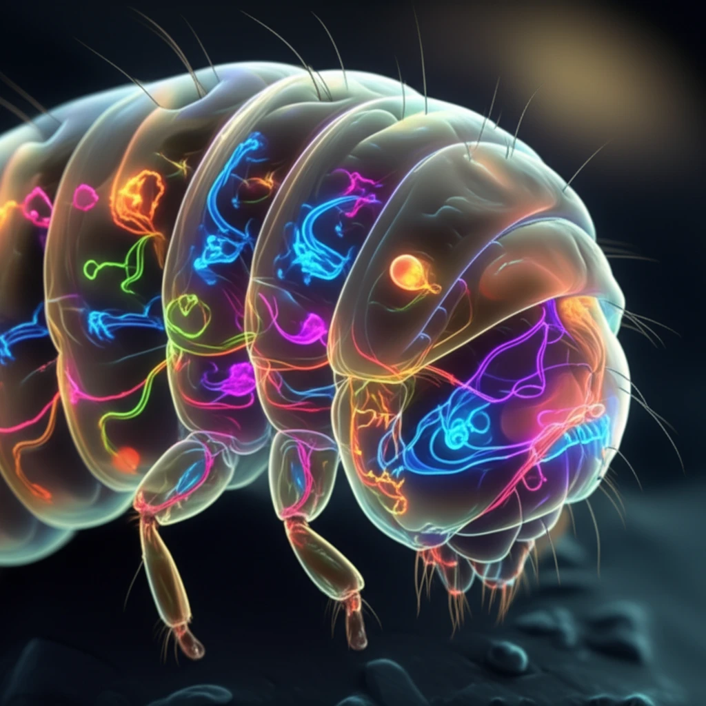
Decoding the Brain: How Scientists Analyze Sensory Neurons in Tiny Fruit Fly Larvae

"Unlocking the Secrets of the Nervous System: A Look at Innovative Techniques for Studying Sensory Neuron Development"
The human brain, a marvel of biological engineering, continues to fascinate and challenge scientists worldwide. Its intricate network of neurons and the complex ways they communicate are fundamental to understanding how we perceive the world, make decisions, and even feel emotions. However, studying the brain is a daunting task due to its size and complexity. This is where innovative research, like that conducted on the humble fruit fly larva, comes into play.
Fruit fly larvae, or Drosophila, might seem an unlikely source of insight into the human brain, but their simple nervous systems offer a unique opportunity for study. Researchers have developed sophisticated methods to analyze the sensory neurons in these tiny creatures, providing valuable clues about how nervous systems develop and function. This research not only advances our understanding of these fundamental biological processes but also paves the way for potential insights into neurological disorders.
This article dives into a fascinating study that investigates the sensory neurons in fruit fly larvae. We'll explore the innovative techniques used to analyze these neurons, the significance of the findings, and how this research contributes to a broader understanding of the nervous system. Join us as we uncover the secrets hidden within these tiny creatures and their potential impact on human health.
Why Sensory Neurons Matter
Sensory neurons are central to how animals detect conditions inside and outside the body. Research on these cells can help clarify how sensory information is converted into neural signals and how those signals influence behavior and physiology. Because findings from tiny fruit fly larvae may not transfer directly to humans, their broader significance should be interpreted cautiously.
Methods and Limits
Scientists commonly combine microscopy, genetic labeling, physiological recording, and molecular analysis to study sensory neurons. These methods can reveal neuronal structure, activity, and gene expression, but each captures only part of the underlying biology. Results may also be limited by experimental conditions and by differences between larval models and more complex nervous systems.
From Microscopes to Neurons
The history of nervous-system research extends from early observations to the recognition of neurons as specialized electrical signaling cells in multicellular animals. Christian Gottfried Ehrenberg made numerous microscopic observations of nerves and ganglia, including the human vagus nerve, contributing to the early visual record of sensory anatomy. Later work gradually established that vagal sensory neurons help monitor internal-organ physiology and maintain homeostasis, making them important mediators of brain-body communication. This historical progression illustrates how anatomical observation laid groundwork for modern sensory-neuron research.
Unveiling the Secrets: The Role of Sensory Neurons and Genetic Mosaics
Sensory neurons are the gatekeepers of our senses, detecting signals from the environment and relaying them to the brain. In fruit fly larvae, these neurons are responsible for sensing touch, temperature, and other stimuli, allowing them to navigate their surroundings and respond to threats. Understanding how these neurons develop and function is key to understanding the broader mechanisms of the nervous system. The study described in this article focuses on a specific type of sensory neuron called dendritic arborization (DA) neurons.
- MARCM (Mosaic Analysis with a Repressible Cell Marker): This technique allows researchers to label individual neurons, making it easier to study their structure and function.
- Flp-out: Another technique that helps to create a 'mosaic' of cells, allowing for detailed analysis of individual neurons within the complex nervous system.
- In vivo imaging: Using advanced microscopy techniques, scientists can observe the neurons in their natural environment.
- Immunohistochemistry: This technique uses antibodies to stain specific proteins within the neurons, revealing their detailed structure.
Current Research Directions
Recent research on sensory neurons is increasingly combining structural, physiological, and molecular approaches. Studies of small model organisms can provide controlled settings for examining how sensory circuits develop and respond to stimuli. However, the latest findings should be treated as an evolving research picture rather than as a complete account of sensory processing.
Limits of the Model
No single method can fully capture how sensory neurons detect stimuli, communicate with other cells, and shape behavior. A result observed in a tiny fruit fly larva may depend on the organism's developmental stage or experimental environment. For that reason, apparent failures or contradictions may reflect differences in measurement and biological context rather than a simple failure of the underlying research question.
Sensory Versus Motor Neurons
Sensory neurons are afferent cells that relay information from the periphery toward the brain or central nervous system, whereas motor neurons are efferent cells that carry commands outward. Reviews comparing sensory and motor neurons emphasize that their regenerative responses differ, with sensory neurons often discussed through work on dorsal root ganglion neurons. Sensory and non-sensory neurons can also use cell-type-specific alternative-splicing programs, showing that neuronal identity involves more than a simple difference in signal direction. These comparisons help place sensory-neuron findings in a broader framework of neuronal specialization.
The Future of Neuroscience: What This Research Means for Us
The study of sensory neurons in fruit fly larvae provides a foundation for understanding the development and function of nervous systems. The techniques and insights gained from this research can be applied to studies of more complex organisms, including humans. This knowledge has the potential to help us understand neurological disorders, develop new treatments, and even enhance our understanding of consciousness. As research continues to advance, we can look forward to more breakthroughs that will unlock the secrets of the brain and improve human health.
A Broader Sensory-Neuron Picture
Recent work presents sensory neurons as more than passive detectors of external stimuli. A 2025 PLOS One study reported a method for extracting and comprehensively analyzing the surfaceome of human sensory-like neurons, creating a framework for studying cellular disease mechanisms. Reviews published in 2024 and 2025 describe sensory neurons as participants in tissue homeostasis, pathogen detection, immune modulation, and behavioral reflexes. Together, these findings suggest that sensory-neuron analysis connects neural signaling with cellular and immune biology.
What Comes Next
Future studies will likely seek to connect neuronal structure, activity, gene expression, and behavior in the same experimental systems. Improved tools may make it easier to compare observations from fruit fly larvae with findings from other organisms. Such work could clarify which mechanisms are broadly shared and which are specific to particular species or developmental stages.
Connecting Levels of Biology
Sensory-neuron research spans multiple levels, from molecular features and cellular circuits to behavior and organism-wide physiology. Integrating these levels remains challenging because different experiments measure different aspects of neural function. Careful replication, transparent methods, and comparisons across models will be important for building conclusions that extend beyond a single laboratory system.
Why the Research Matters
Understanding sensory neurons may eventually inform research on neurological, sensory, and immune-related disorders. Model organisms cannot reproduce every feature of human biology, but they can provide tractable systems for testing mechanisms and methods. The practical value of this work therefore depends on combining discoveries from simple models with validation in human cells and tissues.